
The Cell Cycle
E N D
Presentation Transcript
The Cell Cycle Dr.rer.nat., Dra. Asmarinah, MS Depart of Medical Biology Faculty of Medicine University of Indonesia
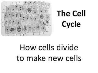
Introduction ■ “Cell doctrine”:where a cell arise, there must be a previous cell (Virchow, 1858) ■ Cell reproduce to deliver message for the continuity of life Cell cycle The cycle that a cell reproduces by an orderly sequence of events in which it duplicates its content and then divides in two
Cell cycles in vivo • Depends on the capacity to divide, there are 3 categories of cells: • Cells that lack the ability to divide and are highly specialized, Examples: nerve cells, muscle cells or red blood cells • Cells that normally don’t divide but can be induced to divide when given an appropriate stimulus. Examples: liver cells, lymphocytes • Cells that normally posses a relatively high level of mitotic activity. Examples: spermatogonia, hematopoietic stem cells and cells at the base of the epithelia that line the body cavities and the body surface
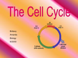
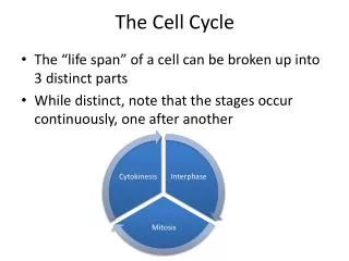
2 main stages in the cell cycle: • Chromosome/DNA replication or duplication • Occur during S phase (S = synthesis) • Chromosome segregation • Occur in M phase (M = mitosis) Between these phase there are G phase (G = Gap) which in the cell require more time to monitor the environment and to grow and double their mass G1: Between M phase and S phase to monitor the internal and external environment to ensure that conditions are suitable and preparations are complete for entering the next phase G2: Between S phase dan M phase to grow and double their proteins and organelles before the entering M phase
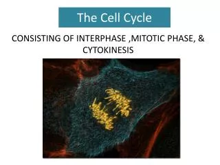
Cell cycle is divided into 4 sequential phases : • M phase, consist of: • * Kariokinesis: nuclear division • * Cytokinesis: cytoplasmic division • - G1 phase • - S phase interphase • - G2 phase Some features of the cell cycle including the time required to complete certain events, vary greatly from one cell to another, even in the same organism. The basic organization of the cell cycle and its control system are essentially the same in all eucaryotic cell
Cell cycle and division Cell cycle
Methods for determination of the cell cycle phase • 1. Microscope: • determine phases in the mitosis (kariokinesis and cytokinesis). • Autoradiographic technique: • 3H-tymidin dan Bromo-deoksiuridin (BrdU) can be incorporated into newly • synthesized DNA detect S phase • DNA-binding fluorescent dyes and a “flow cytometer” • By measuring of DNA content asses the stage that a cell has reached • in the cell cycle and determine the length of G1, S and G2 + M phases
Cell cycle control system • to respon and to monitor the internal and eksternal environment • for the continuing of the cell cycle. • - Play a role in the regulation of the amount of the cell in tissue • To trigger and control the major process of the cell cycle, i.e: • DNA replication, nuclear and cytoplasmic division • “checkpoint “ in cell cycle
Control system in the cell cycle The control system can arrest the cell cycle at specific checkpoints
3 checkpoint system in the cell cycle • G1 “checkpoint” • Occur at the end of the G1 phase before entering S phase • G2 “checkpoint” • at the end of G2 phase before M phase • 3. Metafase “checkpoint” • at the end of metaphase in mitosis to enter anaphase.
2 key components of the cell cycle control system 1. Cyclins will be synthezed dan destroyed in each cell cycle 2. “Cyclin-dependent kinase” (CdK) Protein kinases that its activity rise and falls during the cell cycle. CdK level are constant in the cell cycle Without Cyclin, CdK enzyme inactive
Combination of two key component in the control system of the cell cycle
4 combination of the cyclin and CdK in the cell cycle and their function 1. G1-cyclin (cyclin D) + CdK = G1-CdK helps promote passage through Start or the restriction point in late G1. 2. G1/S-cyclin (cyclin E) + CdK = G1/S-CdK Commit the cell to DNA replication . 3. S-cyclin (cyclin A) + CdK = S-Cdk required for the initiation of DNA replication. production DNA polimerase The activity of S-CdK still high during G2 and begin M prevent rereplication
Continued • M-cyclin (cyclin B) + Cdk = M-Cdk • M-Cdk promote the event of mitosis, i.e: • - phosphorilation of kondensin to changes of the DNA coiling in • chromosom condensation process • - phosphorilation of lamin, to digest of nuclear lamina for the breaking • of nuclear membrane, • - phosphorilation of kinesin, play a role in the formation and the function • of spindel fiber; as well as katastropin and • microtubule-associated protein for the stabilization of microtubule • - activation of APC (“Anaphase Promoting Complex), for digestion of • kohesin in the chromosome segregation process
Cyclin – CdK inhibitor complexs • Cdk Inhibitor protein (CKI) • Exp: Protein 27 (p27) to control G1 and S phase • Protein Wee1 • phosphorilate the active site of CdK enzyme
DNA damage checkpont in the cell cycle • At the end of G1 phase • prevent entry into S phase. DNA damage leads to the activation of the gene regulatory protein p53 which stimulates the trancription of several genes such as p21. This protein inhibits the activity of G1/S-CdK dan S-CdK. • At the end of G2 phase • DNA damage inactivate the phosphatase CDc25 that it can blocks the phosphorilation and activation of M-CdK, thereby blocking entry into mitosis. • When the DNA damage is repaired, the inhibitory signal is turned off, and cell-cycle progression resumes.
“DNA damage checkpoint” At the end of G1 phase
Cell division • Bacteria • Compared to the complex steps of mitosis, the process of cell division of bacteria is much simpler. • Sometime early in the bacteria's life, a second copy of its DNA is made. At the replication origin, which is a specific site on the chromosome. The two copies are attached side by side to the cell membrane. • Cell division of bacteria called binary fission. Binary fission is division in which the cell pinches itself in two, creating two equal or nearly equal halves.
Sequential events of cell division in bacteria: • - Between the two attached DNA genomes, a new plasma membrane and cell wall components are built. • As new materials continue to be added on, the cell is slowly pinched in two by the plasma membrane, pushing inward. Because the location where the cell constriction starts is between the two DNA copies, each daughter cell is ensured one copy. • - the cell is divided and a new cell wall forms around the new membrane, creating two cells from one.
Eukaryotic cell • Consist of two events, i.e: • Nuclear division (kariokinesis) • - Profase • - Prometafase • - Metafase • - Anafase • - Telofase • Cytoplasmic division (cytokinesis) • If it doesn’t happened, leads multinucleated cell.
Interphase • During interphase, • increasing of the cell size • DNA replication • Duplication of the centrosome
Prophase • Replicated chromosome that contains two closely associated sister chromatid, condense • the mitotic spindle assambles between the two centrosome which have replicated and moved to apart in the outside of nucleus • Nuclear membrane still intake
Prometaphase • - Breakdown of the nuclear envelope • Chromosomes can attach to spindle microtubule via their kinetochores and undergo active movement
Metafase • The chromosomes are aligned at the equator of the spindle, midway between the spindle poles. • Kinetochore microtubules attach sister chromatids to opposite poles of the spindle
Anaphase • The sister chromatid synchronously separate to form two daugther chromosomes, and each is pulled slowly towards the spindle pole it face. • Kinetochore microtubules get shorter, and the spindle poles also move apart; both processes contribute to the chromosome separation
Telophase • The two sets of daughter chromosomes arrive at the poles of the spindle and decondense • A new nuclear envelope reassembles around each set, completing the formation of two nuclei and marking the end of mitosis • The division of the cytoplasm begins with the assembly of the contractile ring.
Cytokinesis • The cytoplasm is divided in two by a contractile ring of actin and myosin filament, which pinches the cell in two to create two daughters, each with one nucleus
Meiosis • = “reduction” (Greek word) • ensure of production of haploid phase in the life cycle • Divided into: • * meiotic division I : each chromosome (consisting of two chromatid) is separated from its homologue • * meiotic division II : two chromatid of each chromosome are separated from one another • - By mixing maternal and paternal (recombinant) allele between homologue chromosomes at the profase I, result in increasing of the genetic variability of organism from one generation to the next, with novel ge
Spermatogenesis Oogenesis
Leptotene: chromosome become visible in the light microskope. In the electrone microscope, chromosome are revealed to be composed of paired chromatids • Zygotene: is marked by the visible association of homologous with one another. This process of chromosome pairing is called by synapsis • Pachytene: is caharacterized by a fully formed synaptonemal complex (SC), i.e: a ladder-like structure with tranverse protein filament connecting the two lateral element • Diplotene: is recognized by the dissolution of SC, which leaves the chromosome attached to one another at specific point, termed chiasmata • Diakinesis: the meiotic spindle is assembled and the chromosome are prepared for separation
Extracellular control of cell division, cell growth, and apoptosis • Mitogen, stimulate cell division that otherwise block progress through the cell cycle. • Growth factor, stimulate cell growth (an increase in cell mass) by promoting the synthesis of proteins and other macromolecules and by inhibiting their degradation • Survival factor, promote cell survival by suppresing apoptosis
APOPTOSIS • Programmed cell death (= “falling off” as leaves from a tree) • Homeostasis the number of cells in the community is tightly regulated by controlling the rate of cell death For examples: In a healthy adult human, billions of cells die in the bone marrow and intestine every hour
Ultrastructural Changes during Apoptosis Apoptotic body phagocytosis Nuclear condensation Swelling in plasma membrane The cell is wrinkly Chromatid condensation and fragmentation phagocyte cell
APOPTOSIS Cell is wrinkly Fragmentation plasma membrane intake Cell contents are present No inflamation reaction APOPTOSIS vs NECROSIS NECROSIS • Cell is swelling • Disintegration • Plasma membran destroyed • Cell contents go to the outside • Inflamation reaction
References: Albert et al., Molecular Biology of the Cell. Garland Scientific. 4th ed. 2002. Karp G. Cell and Molecular Biology. 4th ed. 2005 Lodish et al, Molecular Biology of the Cell. 4th ed. 2000