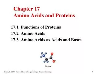
Chapter 17 Amino Acids and Proteins
1.01k likes | 1.82k Vues
Chapter 17 Amino Acids and Proteins. 17.1 Functions of Proteins 17.2 Amino Acids 17.3 Amino Acids as Acids and Bases. Functions of Proteins. Proteins perform many different functions. 17.3 Amino Acids. Amino acids : Are the building blocks of proteins.

Chapter 17 Amino Acids and Proteins
E N D
Presentation Transcript
Chapter 17 Amino Acids and Proteins 17.1 Functions of Proteins 17.2 Amino Acids 17.3 Amino Acids as Acids and Bases
Functions of Proteins Proteins perform many different functions.
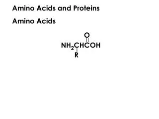
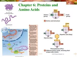
17.3 Amino Acids Amino acids: • Are the building blocks of proteins. • Contain a carboxylic acid group and an amino group on the alpha () carbon. • Have different side groups R that give each amino acid unique characteristics. R side chain | H2N—C —COOH General structure of an | -amino acid H
Nonpolar Amino Acids • Amino acids are classified as nonpolar when the R groups are H, alkyl, or aromatic.
Polar Amino Acids • Amino acids are classified as polar when the R groups are alcohols, thiols, or amides.
Acidic and Basic Amino Acids • Amino acids are classified as acidic when the R group is a carboxylic acid. • Amino acids are classified as basic when the R group is an amine.
17.2 Chiral Objects • Chiral compounds have the same number of atoms arranged differently in space. • A chiral carbon atom is bonded to four different groups. • Your hands are chiral. Try to superimpose your thumbs, palms, back of hands, and little fingers.
Mirror Images • The mirror images of chiral compounds cannot be superimposed. • When the H and I atoms are aligned, the Cl and Br atoms are on opposite sides.
Achiral Structures are Superimposable • When the mirror image of an achiral structure is rotated, the structure can be aligned with the initial structure. Thus this mirror image is superimposable.
Learning Check Identify each as a chiral or achiral compound.
Solution Identify each as a chiral or achiral compound. Chiral Achiral Chiral
Zwitterions • Both the –NH2 and the –COOH groups in an amino acid undergo ionization in water. • A zwitterion forms that has + and – charge. • At the isoelectric point (pI), the + and – charges in the zwitterion are equal. + NH2—CH2—COOH H3N—CH2—COO– Glycine Zwitterion of glycine
Amino Acids as Acids • In solutions more basic than the pI, the —NH3+ in the amino acid donates a proton. + OH– H3N—CH2—COO–H2N—CH2—COO– Zwitterion Negative ion at pI Higher pH
Amino Acids as Bases • In solution more acidic than the pI, the COO- in the amino acid accepts a proton. + H+ + H3N—CH2—COO– H3N—CH2—COOH Zwitterion Positive ion at pI Low pH
pH and Ionization • Acidic amino acids such as aspartic acid have a second carboxyl group that can donate and accept protons. • The pI for aspartic acid occurs at a pH of 2.8.
Electrophoresis • Electrophoresis separates amino acids according to their isoelectric points. • The positively charged amino acids move towards the negative electrode. • The negatively charged amino acids move toward the positive electrode. • An amino acid at its pI will not migrate in either direction.
Separation of Amino Acids • When electrophoresis is completed, the amino acids are identified as separate bands on the filter paper or thin layer plate.
Learning Check CH3 CH3 +|| H3N—CH—COOH H2N—CH2—COO– (1) (2) Which structure represents: A. Alanine at a pH above its pI? B. Alanine at a pH below its pI?
Solution CH3 CH3 +|| H3N—CH—COOH H2N—CH2—COO– (1) (2) Which structure represents: A. Alanine at a pH above its pI? (2) B. Alanine at a pH below its pI? (1)
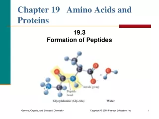
The Peptide Bond • A peptide bondis an amide bond between the carboxyl group of one amino acid and the amino group of the next amino acid. O CH3 O +||+ | || H3N—CH2—C—OH + H3N—CH—C—O– O H CH3 O +|||| || H3N—CH2—C—N—CH—C—O– peptide bond
A Dipeptide • A peptide is named from the free amine (NH3+) using -yl endings for the names of the amino acids. • The last amino acid with the free carboxyl group (COO-) uses its amino acid name.
Learning Check Write the names and three-letter abbreviations of the amino acids in the tripeptides that could form from two glycine and one alanine.
Solution Write the names and three-letter abbreviations of the amino acids in the tripeptides that could form from two glycine and one alanine. Gly-Gly-Ala Glycylglycylalanine Gly-Ala-Gly Glycylalanylglycine Ala-Gly-Gly Alanylglycylglycine
Learning Check Write the name of the following tetrapeptide using amino acid names and three-letter abbreviations.
Solution Ala-Leu-Cys-Met Alanylleucylcysteinylmethionine Ala Leu Cys Met
17.7 Primary Structure • A polypeptide containing 50 or more amino acids is called a protein. • The primary structure of a protein is the sequence of amino acids in the peptide chain. Ala-Leu-Cys-Met
Primary Structures • The nonapeptides oxytocin and vasopressin have similar primary structures. • Only the amino acids at positions 3 and 8 differ.
Insulin Insulin: • Was the first protein to have its primary structure determined. • Of humans has a primary structure that is similar to the insulin of pigs and cows.
17.9 Secondary Structure: Alpha Helix The secondary structures of proteins indicate the arrangement of the polypeptide chains in space. • The alpha helix is a three-dimensional arrangement of the polypeptide chain that gives a corkscrew shape like a coiled telephone cord.
Alpha Helix • The coiled shape of the alpha helix is held in place by hydrogen bonds between the amide groups and the carbonyl groups of the amino acids along the chain.
Secondary Structure: Pleated Sheet The pleated sheet: • Holds proteins in a parallel arrangement with hydrogen bonds. • Has R groups that extend above and below the sheet. • Is typical of fibrous proteins such as silk.
Secondary Structure: Triple Helix A triple helix: • Consists of three alpha helix chains. • Contains large amounts glycine, proline, hydroxy proline and hydroxylysine that contain –OH groups for hydrogen bonding. • Is found in collagen, connective tissue, skin, tendons, and cartilage.
Essential Amino Acids Essential amino acids: • Are the ten amino acids that are not synthesized by the body. • Must be obtained from the diet.
Essential Amino Acids Essential amino acidsare: • Found in milk and eggs (complete proteins). • Not all found in grains and vegetables (incomplete proteins). • Obtained by combining two or more vegetables that provide complementary proteins.
Learning Check Indicate the type of structure as: 1) primary 2) alpha helix 3) beta pleated sheet 4) triple helix A. Polypeptide chains held side by side by H bonds. B. Sequence of amino acids in a polypeptide chain. C. Corkscrew shape with H bonds between aminoacids. D. Three peptide chains woven like a rope.
Solution Indicate the type of structure as: 1) primary 2) alpha helix 3) beta pleated sheet 4) triple helix A. 3 Polypeptide chains held side by side by H bonds. B. 1 Sequence of amino acids in a polypeptide chain. C. 2 Corkscrew shape with H bonds between amino acids. D. 4 Three peptide chains woven like a rope.
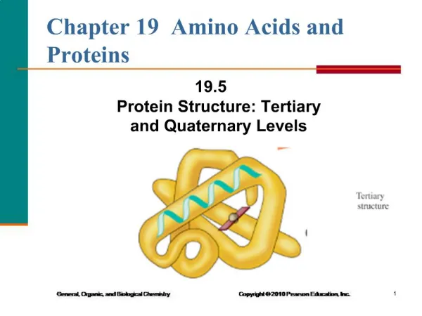
17.10 Tertiary Structure The tertiary structure: • Gives a specific overall shape to a protein. • Involves interactions and cross links between different parts of the peptide chain. • Is stabilized by Hydrophobic and hydrophilic interactions Salt bridges Hydrogen bonds Disulfide bonds
Tertiary Structure • The interactions of the R groups give a protein its specific three-dimensional tertiary structure.
Globular Proteins Globular proteins: • Have compact, spherical shapes. • Carry out synthesis, transport, and metabolism in the cells. • Such as myoglobin store and transport oxygen in muscle. Myoglobin
Fibrous Proteins Fibrous proteins: • Consist of long, fiber-like shapes. • Such as alpha keratins make up hair, wool, skin, and nails. • Such as feathers contain beta keratins with large amounts of beta-pleated sheet structures.
Learning check Select the type of tertiary interaction as: 1) disulfide 2) ionic 3) H bonds 4) hydrophobic A. Leucine and valine B. Two cysteines C. Aspartic acid and lysine D. Serine and threonine
Solution Select the type of tertiary interaction as: 1) disulfide 2) ionic 3) H bonds 4) hydrophobic A. 4 Leucine and valine B. 1 Two cysteines C. 2 Aspartic acid and lysine D. 3 Serine and threonine
17.11 Quaternary Structure • The quaternary structure contains two or more tertiary subunits. • Hemoglobin contains two alpha chains and two beta chains. • The heme group in each subunit picks up oxygen for transport in the blood to the tissues.
Learning Check Identify the level of protein structure as: 1) Primary 2) Secondary 3) Tertiary 4) Quaternary A. Beta pleated sheet B. Order of amino acids in a protein C. A protein with two or more peptide chains D. The shape of a globular protein E. Disulfide bonds between R groups
Solution Identify the level of protein structure 1. Primary 2. Secondary 3. Tertiary 4. Quaternary A. 2 Beta pleated sheet B. 1 Order of amino acids in a protein C.4 A protein with two or more peptide chains D. 3 The shape of a globular protein E. 3 Disulfide bonds between R groups
17.12 Chemical Properties of Proteins Protein hydrolysis: • Splits the peptide bonds to give smaller peptides and amino acids. • Occurs in the digestion of proteins. • Occurs in cells when amino acids are needed to synthesize new proteins andrepair tissues.