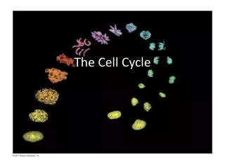
The Cell Cycle
The Cell Cycle. Key Concepts. Most division results in genetically identical cells Cell cycle consists of alternating periods of mitosis and interphase Eukaryotic cell cycle is highly regulated. Purpose. Ultimate purpose of cell cycle is to propagate genetic information Also important for

The Cell Cycle
E N D
Presentation Transcript
Key Concepts • Most division results in genetically identical cells • Cell cycle consists of alternating periods of mitosis and interphase • Eukaryotic cell cycle is highly regulated
Purpose • Ultimate purpose of cell cycle is to propagate genetic information • Also important for • Tissue repair • Growth • Reproduction
ChromosomalDNA molecules Figure 12.5-1 Chromosomes Centromere 1 Chromosomearm
ChromosomalDNA molecules Figure 12.5-2 Chromosomes Centromere 1 Chromosomearm Chromosome duplication(including DNA replication)and condensation 2 Sisterchromatids
ChromosomalDNA molecules Figure 12.5-3 Chromosomes Centromere 1 Chromosomearm Chromosome duplication(including DNA replication)and condensation 2 Sisterchromatids Separation of sisterchromatids intotwo chromosomes 3
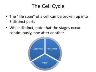
Phases of cell cycle • Interphase • G1 • G2 • S • Mitosis • Prophase • Metaphase • Anaphase • Telophase
Figure 12.6 INTERPHASE S(DNA synthesis) G1 Cytokinesis G2 Mitosis MITOTIC(M) PHASE
Figure 12.7 10 m G2 of Interphase Prophase Prometaphase Metaphase Anaphase Telophase and Cytokinesis Centrosomes(with centriole pairs) Chromatin(duplicated) Fragments of nuclearenvelope Nonkinetochoremicrotubules Early mitoticspindle Aster Metaphase plate Cleavagefurrow Nucleolusforming Centromere Plasmamembrane Nuclearenvelope Chromosome, consistingof two sister chromatids Kinetochore Kinetochoremicrotubule Nucleolus Nuclearenvelopeforming Daughterchromosomes Centrosome atone spindle pole Spindle
Figure 12.7a Prometaphase G2 of Interphase Prophase Fragments of nuclearenvelope Centrosomes(with centriole pairs) Early mitoticspindle Nonkinetochoremicrotubules Chromatin(duplicated) Aster Centromere Plasmamembrane Kinetochore Nucleolus Kinetochoremicrotubule Chromosome, consistingof two sister chromatids Nuclearenvelope
Figure 12.7b Metaphase Anaphase Telophase and Cytokinesis Nucleolusforming Metaphase plate Cleavagefurrow Nuclearenvelopeforming Spindle Centrosome atone spindle pole Daughterchromosomes
Mitotic Spindle Centrosome Aster Metaphaseplate(imaginary) Sisterchromatids Microtubules Chromosomes Kineto-chores Centrosome 1 m Overlappingnonkinetochoremicrotubules Kinetochoremicrotubules 0.5 m
EXPERIMENT Figure 12.9a Kinetochore Spindlepole Mark RESULTS
Figure 12.9b CONCLUSION Chromosomemovement Kinetochore Microtubule Tubulinsubunits Motor protein Chromosome
(a) Cleavage of an animal cell (SEM) Figure 12.10a 100 m Cleavage furrow Daughter cells Contractile ring ofmicrofilaments
(b) Cell plate formation in a plant cell (TEM) Figure 12.10b Vesiclesformingcell plate Wall of parent cell 1 m New cell wall Cell plate Daughter cells
Binary Fission • Prokaryotes replicate via binary fission • Genome replication starts at location called origin of replication • Cell membrane pinches around midline of cell
Cell wall Origin ofreplication Figure 12.12-1 Plasma membrane E. coli cell Bacterial chromosome 1 Chromosomereplicationbegins. Two copies of origin
Cell wall Origin ofreplication Figure 12.12-2 Plasma membrane E. coli cell Bacterial chromosome 1 Chromosomereplicationbegins. Two copies of origin 2 Origin Origin Replicationcontinues.
Cell wall Origin ofreplication Figure 12.12-3 Plasma membrane E. coli cell Bacterial chromosome 1 Chromosomereplicationbegins. Two copies of origin 2 Origin Origin Replicationcontinues. 3 Replicationfinishes.
Cell wall Origin ofreplication Figure 12.12-4 Plasma membrane E. coli cell Bacterial chromosome 1 Chromosomereplicationbegins. Two copies of origin 2 Origin Origin Replicationcontinues. 3 Replicationfinishes. 4 Two daughtercells result.
Evolution of mitosis • Eukaryotes most likely evolved from prokaryotic organisms • Some organisms display intermediate “levels” of mitotic behavior.
Figure 12.15 G1 checkpoint Cell Cycle Regulation Controlsystem S G1 G2 M M checkpoint G2 checkpoint
Cyclins and CDKs • Two types of regulatory proteins in cell cycle control: cyclins and cyclin-dependent kinases (Cdks) • Cdks activity fluctuates during cell cycle because it is controled by cyclins, so named because their concentrations vary with the cell cycle • MPF (maturation-promoting factor) is a cyclin-Cdk complex that triggers a cell’s passage past the G2 checkpoint into the M phase
Figure 12.17a M M G1 G2 G1 G2 M S G1 S MPF activity Cyclinconcentration Time (a) Fluctuation of MPF activity and cyclin concentration during the cell cycle
Figure 12.17b G1 S Cdk Cyclin accumulation M G2 Degradedcyclin G2checkpoint Cdk Cyclin isdegraded Cyclin MPF (b) Molecular mechanisms that help regulate the cell cycle
Stop and Go Signals • An example of an internal signal is that kinetochores not attached to spindle microtubules send a molecular signal that delays anaphase • Some external signals are growth factors, proteins released by certain cells that stimulate other cells to divide • For example, platelet-derived growth factor (PDGF) stimulates the division of human fibroblast cells in culture
Figure 12.18 Scalpels 1 A sample of humanconnective tissue iscut up into smallpieces. Petridish 2 Enzymes digestthe extracellularmatrix, resulting ina suspension offree fibroblasts. 10 m 4 PDGF is addedto half thevessels. 3 Cells are transferred toculture vessels. With PDGF Without PDGF
External Signals • A clear example of external signals is density-dependent inhibition, in which crowded cells stop dividing • Most animal cells also exhibit anchorage dependence, in which they must be attached to a substratum in order to divide • Cancer cells exhibit neither density-dependent inhibition nor anchorage dependence
Cancer • Normal cells undergo a transformation • Typically result of genetic mutation • Loss of ability to govern cell cycle • Too much growth • Not enough death • Cancers typically pick up more and more mutations as they progress.