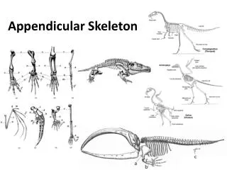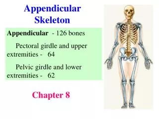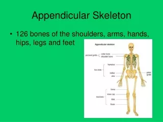
Overview of the Lower Appendicular Skeleton: The Pelvic Girdle and Lower Limb Bones
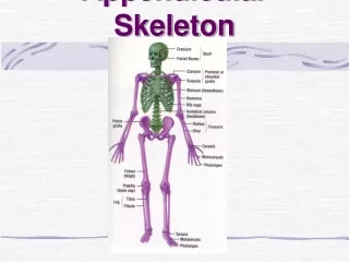
The lower appendicular skeleton comprises the pelvic girdle, including the sacrum, coccyx, and the two coxae (hip bones). Each coxa consists of three parts: the ilium, ischium, and pubis, which fuse at the acetabulum. The femur, tibia, fibula, tarsals, metatarsals, and phalanges form the lower limb. The femur is the body's longest bone, while the tibia and fibula contribute to the leg structure. Tarsals include key bones such as the talus and calcaneus, while the foot has metatarsals and phalanges essential for movement and support.

Overview of the Lower Appendicular Skeleton: The Pelvic Girdle and Lower Limb Bones
E N D
Presentation Transcript
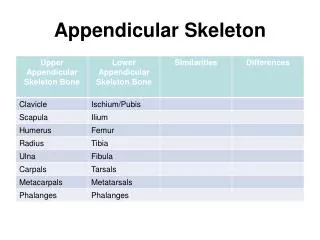
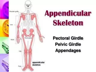
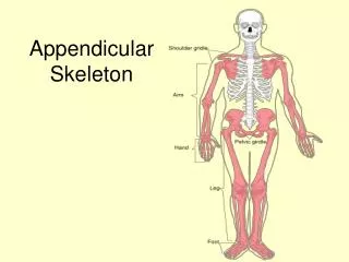
Pelvic Girdle • Composed of sacrum, coccyx, and 2 coxae (hipbones) • Coxae have 3 distinct parts: • Ilium • Ischium • Pubis
Coxae parts fuse together in the acetabulum(acetabul-), a cup-shaped area on the lateral surface of the hip that receives the head of the femur.
Ilium • Largest and uppermost portion of the coxa • The upper edge is called the iliac crest • Joins the sacrum at the sacroiliac joint • Anterior superior iliac spine- the bony prominence you feel as your “hipbone”
Ischium • Forms the lowest portion of the coxa • Ischialtuberosity • Points posteriorly AND downward • Supports the weight of the body when sitting • Ischial spine – a sharp projection above the ischialtuberosity, near the junction of the ischium and ilium
Pubis • Anterior portion of the coxa • Two pubic bones join midline at the symphysis pubis joint • Pubic arch • Angle formed by pubic bones below the symphysis pubis • Arch is wider in females
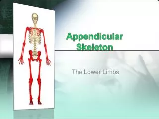
Lower Limb • Femur • Tibia • Fibula • Tarsals • Metatarsals • Phalanges
Femur • Longest and strongest bone in the body • Head at top fits into __________of coxa • Greater trochanter – superior, lateral process • Lesser trochanter – inferior, medial process • Distal end: • Two rounded processes posteriorly: lateral condyleand medial condyle • Patella articulates anteriorly
Tibia • aka, “shin bone” • Proximal end: • Medial and lateral condyles are concave and articulate with condyles of the femur • Tibialtuberosity just below the condyles; attachment point for patellar ligament • Distal end: medial malleolusforms prominent bony point of inner ankle
Fibula • Proximal: head • Articulates with tibia just below the lateral condyle • DOES NOT enter into knee joint or bear any weight • Distal: lateral malleolus forms outer prominent bony part of ankle
Ankle “Tarsals” • “Tiger Cubs Need MILC” • Talus Calcaneus (“heel bone”) Navicular Medial cuneiform Intermediate cuneiform Lateral cuneiform Cuboid
“MILC: Need The Calcium” • 1=Medical Cuneiform • 2=Intermediate cuneiform • 3=Lateral cuneiform • 4= Cuboid • 5= Navicular • 6= Talus • 7= calcaneus
Foot • 5 metatarsals • numbered 1-5 starting medially • Heads at distal ends form the ball of the foot • Phalanges • Toes • Each toe has 3 phalanges, except the big toe • What are the phalanges of each toe called? (HINT: Just like the fingers) • Which phalanx is the big toe missing?
https://resources.oncourse.iu.edu/access/content/group/FA09-KO-OTHR-PRAC-18273/Helpful%20Websites/Nicole_s%20Website%20Pages/Bones/Foot-tarsals.JPGhttps://resources.oncourse.iu.edu/access/content/group/FA09-KO-OTHR-PRAC-18273/Helpful%20Websites/Nicole_s%20Website%20Pages/Bones/Foot-tarsals.JPG