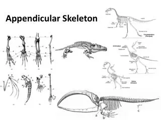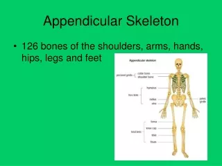
Appendicular Skeleton
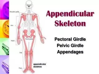
Appendicular Skeleton. Lesson 3. Upper Division: Shoulders and Arms. Shoulder girdle Clavicle: joins the sternum anteriorly and the scapula laterally. Is the most frequently broken bone. Scapula:

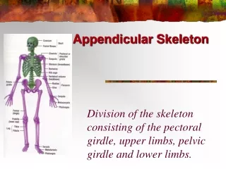
Appendicular Skeleton
E N D
Presentation Transcript
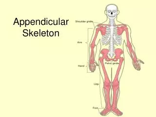
Appendicular Skeleton Lesson 3
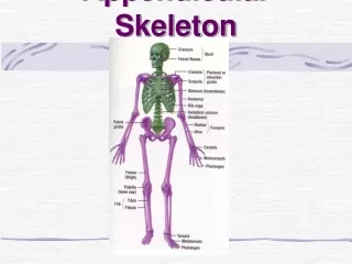
Upper Division: Shoulders and Arms • Shoulder girdle • Clavicle: joins the sternum anteriorly and the scapula laterally. Is the most frequently broken bone. • Scapula: • Spine: the raised ridges; muscles that move the arm attach to the fossae above and below the scapular spine • Acromion: the process that joins the clavicle; it can be felt as the highest point of the shoulder • Glenoid cavity: shallow socket below the acromion; forms a ball-and-socket joint with the humerus • Coracoid process: medial to the glenoid cavity; it serves as a muscle attachment
Upper Extremity • Humerus: the proximal bone; forms a joint with the scapula above and with the radius and the ulna at the elbow • Ulna: lies on the medial side, in line with the little finger • Olecranon: top of the ulna; forms the point of the elbow • Trochlear notch: the semilunar notch, meets the trochlea of the humerus allowing a hinge action at the elbow joint • Radius: lies on the lateral side, above the thumb. When the palm is turned down, the lower end of the radius rotates around the ulna, projecting the styloid process of the ulna at the outside of the wrist.
Wrist: contains eight carpal bones arranged in two rows of four each • Beginning with the thumb: • Row 1: scaphoid, lunate, triquetral, pisiform • Row 2: trapezium, trapezoid, capitate, hamate • Palm: contains five metacarpal bones; their rounded distal ends form the knuckles • Fingers: contain 14 phalanges in each hand; two for the thumb and three for each finger. They are identified by first or proximal, second or middle, and third or distal.
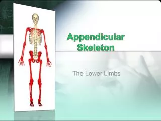
Lower Division: Pelvis and Legs • Pelvic Bones • Pelvic bone: or oscoxae, begins as three separate bones that later fuse • Ilium: forms the upper, flared portion • Iliac crest: curved rim along the upper border of the ilium • Anterior superior iliac spine: most prominent of the bony projections • Ischium: lowest and strongest part of the pelvic bone • Ischial spine: found at the back of the pelvic outlet; used to indicate the progress of childbirth
Ischial tuberosity: below the ischial spine; helps support the weight of the trunk when sitting down • Pubis: forms the anterior part of the pelvic bone; the joint formed is called the pubic symphysis • Portions of all three bones contribute to the formation of the acetabulum, which is the socket for the femur • Obturator foramina: found near the front of each hip bone and partially covered by a membrane • The two ossacoxae join in forming the pelvis. The pelvis supports the trunk and the organs in the lower abdomen.
The Lower Extremity • Femur: the thigh bone; is the longest and strongest bone in the body • Greater trochanter: the large lateral projection at the top • Lesser trochanter: located on the medial side • Patella: the kneecap: embedded in the tendon of the quadriceps femoris where it crosses the knee joint • Tibia: found medially; is the longer, weight-bearing bone of the lower leg. • Medial malleolus: downward projection at the lower end of the tibia; it forms the prominence of the inner aspect of the ankle.
Ankle/foot: contains seven tarsal bones • Heel: calcaneous • Talus: ankle • Three cuniforms that meet the metatarsals • Cuboid: lateral, above the little toe • Instep of foot: contains five metatarsal bones; the heads of these form the ball of the foot • Toes: made up of phalanges; very similar to the fingers in number and identification.
Skeletal Changes in Aging • Vertebral Column • Thinning of intervertebral discs causes a decrease in height around age 40 • Vertebra may also lose height in later years • Costal cartilage becomes calcified and less flexible; chest may decrease in diameter by 1 inch
Joints • Types of Joints • Fibrous joints: held together by fibrous connective tissue • This type of joint is immovable and called a synarthrosis • An example is a skull suture • Cartilaginous joints: bones are connected by cartilage • This type of joint is slightly movable and called an amphiarthrosis • An example is the pubic symphysis • Synovial joints: bones have a potential space between them called the joint cavity that contains synovial fluid, which lubricates the joint • This type of joint is freely movable and called a diarthrosis • Most joints are this type
Structure of Synovial Joints • Held together by ligaments; they reinforce and help stabilize the joints at various points. • Joint capsule: connective tissue that encloses each joint and is continuous with the periosteum of the bones • Articular cartilage: protects the surfaces in freely movable joints • Bursae: small sacs of synovial fluid that help ease movement over and around the joint; inflammation of a bursae is called bursitis
Types of Synovial Joints • Gliding joint: bone surfaces slide over each other; examples include the joints of the wrist and ankle • Hinge joint: allows movement in one direction; examples include the elbow and joints between the phalanges • Pivot joint: allows rotation around the length of the bone; examples include the atlas and axis of the neck • Condyloid joint: allows movement in two directions; examples include the joints between the metacarpals and the phalanges
Saddle joint: allows for more movement than the condyloid joint; an example is the joint between the thumb and the wrist • Ball-and-socket joint: allows movement in all directions around a central point; an example is the shoulder joint
Movement • Flexion: motion that decreases the angle between bones • Extension: motion that increases the angle between bones • Abduction: motion away from the midline of the body • Adduction: motion toward the midline of the body • Circumduction: circle motion around a point • Rotation: twisting or turning a bone on its own axis • Supination: turning the palm up or forward • Pronation: turning the palm down or backward
Inversion: turning the sole inward • Eversion: turning the sole outward • Dorsiflexion: foot bent up at the ankle • Plantar flexion: foot bent down at the ankle