Cell Division and the Cell Cycle
200 likes | 229 Vues
Explore the intricate process of cell division in prokaryotes and eukaryotes, including mitosis and meiosis. Learn about the phases of the cell cycle and the key events that occur during each stage.

Cell Division and the Cell Cycle
E N D
Presentation Transcript
I. Prokaryote Cell Division A. A prokaryote cell divides by binary fission. 1. DNA copies itself. 2. Cell grows and begins to divide. 3. New cell wall forms and two new cells are formed. 4. 2 cells are identical to original.
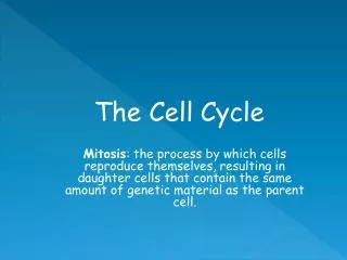
II. Cell Division’s A. Mitosis: body cell division resulting in two cells with identical genetic material 1. 46 chromosomes to 46 B. Meiosis: reproductive cells reducing the number of chromosomes by half from 46 to 23 chromosomes. 1. Also called Reduction Division.
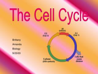
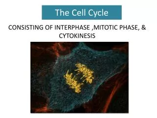
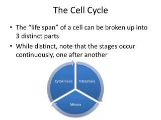
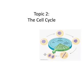
III. The Cell Cycle A. The repeating set of events that make up the life of a cell. 1. Interphase, G1, S, G2 phases 2. M phase: the dividing phase, prophase, metaphase, anaphase, telophase. 3. Cytokinesis: division of cytoplasm of a cell
IV. Interphase: Non-dividing phase A. G1 phase: offspring cells grow to mature size B. S phase: cell’s DNA is copied C. G2 phase: cell prepares for division D. G0 phase: cells not dividing or preparing for division, brain cells.
1. Prophase A. DNA tightens into rod-shaped chromosomes. B. Centromere connects the two sister chromatids C. Nucleolus and nuclear membrane disappears
D. Centrosomes- dark spots that appear next to the disappearing nucleus, containing a pair of cyclinder shaped bodies called centrioles, (animals cells only) E. Spindle fibers radiate from the centromeres and form the mitotic spindle • This equally divides the chromatids.
F. 2 kinds of Spindle Fibers 1. Kinetochore fibers attach to a protein called kinetochore found in the centromere region, and move the chromosomes 2. Polar fibers extend across the dividing cell from one centrosome to the other centrosome.
2. Metaphase A. Kinetochore fibers move the chromosomes to the center of the dividing cell 1). Chromosomes are easily seen lined up in the middle of the cell
3. Anaphase A. Sister chromatids separate at the centromere and move toward opposite ends of the cell 1). They are pulled centromere first, making it look like they are being dragged.
4. Telophase A. Chromosomes are now at opposite ends 1). Spindle fibers disassemble. 2). Chromosomes relax and turn into chromatin. 3). Nuclear envelope and nucleolus reforms.
5. Cytokinesis A. Dividing of the cytoplasm 1). Cleavage furrow forms between the two poles of an animal cell. 2). This pinches the cell into two. 3). In plants, a cell plate or wall forms from vesicles, separating the two halves.