Download
1 / 124
1.25k likes | 1.42k Vues
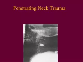
Management of Penetrating Neck Trauma. Ottawa Civic. MVA, aphasia, R hemiplegia. Types of Weapons. Low velocity – knives, ice picks, glass High velocity – handguns, shotguns, shrapnel. K=1/2mv^2. Guns. <. Ballistics. Anatomy. Anatomy. Zone III. Zone II. Zone I.

E N D
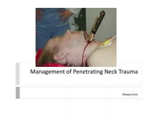
Management of Penetrating Neck Trauma Ottawa Civic
Types of Weapons • Low velocity – knives, ice picks, glass • High velocity – handguns, shotguns, shrapnel K=1/2mv^2
Guns <
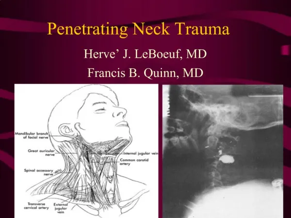
Anatomy Zone III Zone II Zone I
Signs of Injury: Shock, Profuse bleeding, Evolving stroke, Expanding hematoma, hemoptysis, hematemesis, unequal pulses, bruits or thrills Vascular
Signs of Injury: Subcutaneous emphysema, Hoarseness, Respiratory distress, Stridor Larynx/Trachea Neck pain, Blood in saliva, Fever, Odynophagia Esophagus
Initial Management Airway Intubation vs. Surgical Airway Breathing Circulation IV access, Immediate Exploration Examination Determine weapon trajectory
Management of the Stable Patient: The Standard: Wound Penetrates Platysma? No Yes Immediate Neck Exploration Observation/Discharge Laryngoscopy Esophagoscopy
The Standard: • Based on wartime experiences • Fogelman et al (1956) : • immediate neck exploration-> better outcomes in vascular injuries. • negative neck explorations in > 50% • Arteriogram? • screening tool before exploration • zone 1 and 3 injuries • hard to detect on physical • Safe answer on board exam…
Arteriogram • Flint et al (1973): • negative P.E. in 32% of pts. with major zone 1 vascular injury. • Arteriogram can be accompanied by treatment (e.g. embolization).
A Newer Algorithm Mansour et al 1991 retrospective study
Newer Algorithm (Mansour) • 63% of the study population was in the observation group. • Overall mortality 1.5% • similar to those in more rigorous treatment protocols. • Similar results obtained in other large studies with similar protocols (e.g. Biffi et al 1997). • NOTE: Arteriogram in asymptomatic patients with zone 1 injury.
Points of Controversy: • Most trauma surgeons accept observation of select patients similar to the Mansour algorithm. • Study by Eddy et al • questions the necessity for arteriogram / esophagoscopy in asymptomatic zone 1 injury (use of P.E. and CXR resulted in no false negatives). • Other noninvasive modalities than arteriogram exist for screening patients for vascular injury.
CT scan • Can id weapon trajectory and structures • only in stable patients. • Gracias et al (2001) • CT scan in stable patients: • able to save patients from arteriogram indicated by other protocols 50% of the time • avoid esophagoscopy in 90% of tested patients who might otherwise have undergone it.
Duplex Ultrasonography • Requires the presence of reliable technician and radiologist. • A double blinded study by Ginsburg et al (1996) showed 100% true negative, 100% sensitivity in detecting arterial injury, using arteriography as the gold standard.
Management of Vascular Injuries: • Common carotid: • repair preferred over ligation in almost all cases. • Saphenous vein graft may be used. • Shunting is rarely necessary. • Thrombectomy may be necessary. • Internal carotid: • Shunting is usually necessary • Vertebral: • Angiographic embolization • proximal ligation can be used if the contralateral vertebral artery is intact. • Internal Jugular: Repair vs. ligation.
Esophageal Injury: • Diagnosis: • esophagoscopy and esophagram in symptomatic patients. • Injection of air or methylene blue in the mouth may aid in localizing injuries. • Controlled fistula with T-tube • exteriorization of low non-repairable wounds • Small pharyngeal lesions above arytenoids can be treated with NPO and observation 5-7 days • All patients should be NPO for 5-7 days.
Laryngeal/Tracheal Injury • Thorough Direct Laryngoscopy for suspicious wounds • Tracheotomy for suspected laryngeal injury
Thoracic Trauma • 2nd leading cause of trauma deaths • after head injury • 10-20% of all trauma deaths • Many deaths are preventable
Thoracic Trauma • Mechanisms of Injury • Blunt Injury • Deceleration • Compression • Penetrating Injury • Combination
Thoracic Trauma • Anatomical Injuries • Thoracic Cage (Skeletal) • Cardiovascular • Pleural and Pulmonary • Mediastinal • Diaphragmatic • Esophageal • Penetrating Cardiac
Thoracic Trauma • Hypoxia • hypovolemia • pulmonary V/P mismatch • in intrathoracic pressure relationships • Hypercarbia • in intrathoracic pressure relationships • level of consciousness • Impairments to cardiac output • blood loss • increased intrapleural pressures • blood in pericardial sac • myocardial valve damage • Acidosis – final result • hypoperfusion of tissues
Thoracic Trauma • Initial exam directed toward life threatening: • Injuries • Open pneumothorax • Flail chest • Tension pneumothorax • Massive hemothorax • Cardiac tamponade
Thoracic Trauma • Assessment Findings • Mental Status • decreased • Pulse • absent, tachy or brady • BP • narrow PP, hyper- or hypotension, pulsusparadoxus • Ventilatory rate & effort • tachy- or bradypnea, labored, retractions • Skin • diaphoresis, pallor, cyanosis, open injury, ecchymosis
Thoracic Trauma • Assessment Findings • Neck • tracheal position, SQ emph, JVD, open injury • Chest • contusions, tenderness, asymmetry, abN a/e, bowel sounds, abnormal percussion, open injury, impaled object, crepitus, hemoptysis • Heart Sounds • muffled, distant, regurgitantmurmur • Upper abdomen • contusion, open injury
Thoracic Trauma • Assessment Findings • ECG (ST segment abnormalities, dysrhythmias) • History • Dyspnea • Pain • Past hx of cardiorespiratory disease • Restraint devices used • Item/Weapon involved in injury
Thoracic Trauma Specific Injuries
Rib Fracture • MC chest wall injury from direct trauma • More common in adults than children • Especially common in elderly • Most commonly 5th - 9th ribs • Poor protection
Rib Fracture • Fractures of 1st and 2nd second require high force • Frequently have injury to aorta or bronchi • Occur in 90% of patients with tracheo-bronchial rupture • May injure subclavian artery/vein • 30% will die
Rib Fracture • Fractures of 10 to 12th ribs can cause damage to underlying abdominal solid organs: • Liver • Spleen • Kidneys
Rib Fracture • Management • PPV • Analgesics for isolated trauma • Non-circumferential splinting • Monitor elderly and COPD patients closely • Broken ribs can cause decompensation • Patients will fail to breathe deeply and cough, resulting in poor clearance of secretions
Sternal Fracture • Uncommon, 5-8% in blunt chest trauma • Large traumatic force • Direct blow to front of chest by • Deceleration • steering wheel • dashboard • Other object
Sternal Fracture • 25 - 45% mortality due to associated trauma: • Disruption of thoracic aorta • Tracheal or bronchial tear • Diaphragm rupture • Flail chest • Myocardial trauma • High incidence of • myocardial contusion, cardiac tamponade or pulmonary contusion
Sternal Fracture • Management • Establish airway • High concentration oxygen • Assist ventilations as needed • IV NS/LR • Restrict fluids • Rule out associated injuries
Flail Chest • Usually secondary to blunt trauma • Most commonly in MVA • Also results from • falls from heights • industrial accidents • assault • birth trauma • More common in older patients
Flail Chest • Mortality rates 20-40% due to associated injuries • Mortality increased with • advanced age • seven or more rib fractures • three or more associated injuries • shock • head injuries
Flail Chest • Consequences of flail chest • Respiratory failure due to • pulmonary contusion • inadequate diaphragm movement • Paradoxical movement of the chest • must be large to compromise ventilation • Increased work of breathing • decreased chest expansion • pain
Flail Chest • Suspect spinal injuries • Establish airway • Assist ventilation • Treat hypoxia from underlying contusion • Promote full lung expansion • Consider need for intubation and PEEP • Mechanically stabilize chest wall • questionable value
Flail Chest • Management • IV of LR/NS • Avoid rapid replacement in hemodynamically stable patient • Contused lung cannot handle fluid load • Monitor EKG • Chest trauma can cause dysrhythmias