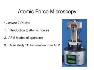

Atomic Force Microscopy
E N D
Presentation Transcript
Atomic Force Microscopy XiaoyuChe
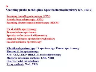
What’s AFM? • Atomic force microscopy(AFM) is one of the foremost tools for imaging, measuring and manipulating matter at the nanoscale • A type of scanning probe microscopy(SPM) • SPM: Forms images of surfaces using a physical probe that scans the specimen • Scanning Tunneling microscope(STM, the predecessor of AFM) is also a type of SPM.
What’s AFM? • High-resolution mapping of surface topography, by far the biggest application of the AFM • Offers image resolution down to the atomic scale
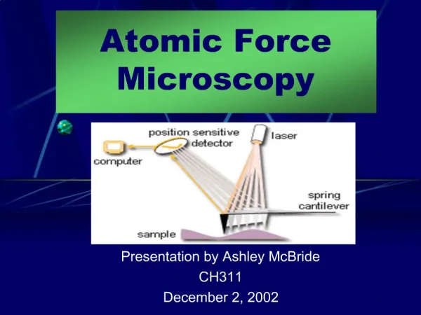
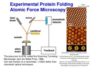
How does aFM work? • The information is gathered by “feeling” the surface with a mechanical probe. • A cantilever with a sharp tip, which is typically silicon or silicon nitride. • The tip radius of curvature is very small, on the order of nanometers to ensure accuracy. So how does it feels the sample?
How does aFM work? Let’s Review Hooke’s law Hooke’s law: The force F needed to extend or compress a spring by some distance x is proportional to that distance. Formula: F=-kx
How does aFM work? • In atomic scale it is not spring-mass system anymore. (contact force, van der Waals force, capillary force, chemical bonding, electrostatic force, magnetic force, etc.) • So when the tip approaches the surface it can “feel” these forces and the deflection is measured to be converted into image information. That’s why it is called atomic force microscopy!
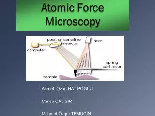
How does aFM work? • But how we measure the deflection? • Use a laser spot reflected from the top surface of the cantilever into an array of photodiodes. • Position sensitive detector(PSD) Angular displacement Differential amplifier Two closely spaced photodiodes
Different imaging mode • Static mode (contact mode) dynamic mode (non-contact mode and tapping mode) • Static mode: • Dynamic mode:
Contact mode • Attractive forces can be quite strong, cause the tip to contact the surface. • Mainly used to image hard surfaces when the presence of lateral forces is not expected to modify the morphological features. • On crystalline surfaces such as mica, Au (111), salt crystals, etc. • Prone to noise and drift • low stiffness cantilevers are used to boost the deflection. • Si probes are more common CdF2 films grown on a CaF2 (111) substrate. Scan is taken in contact mode using a CSC21 probe (now upgraded to HQ:XSC11). Scan size 2 x 2 µm, height 2 nm..
Non-contact mode • The tip of the cantilever does not contact the sample surface. • Oscillated at either its resonant frequency (frequency modulation) or just above (amplitude modulation) • The van der Waals forces or any other long range force acts to decrease the resonance frequency of the cantilever. • Maintains a constant oscillation amplitude or frequency by adjusting the average tip-to-sample distance. • Tip-to-sample distance at each (x,y) data point , construct a topographic image of the sample surface.
Non-contact mode • Does not suffer from tip or sample degradation effects • If a few monolayers of adsorbed fluid are lying on the surface of a rigid sample, the images may look quite different. • Frequency modulation:changes in the oscillation frequency provide information about tip-sample interactions. • Amplitude modulation: changes in the oscillation amplitude or phase provide the feedback signal for imaging
Tapping mode • In ambient conditions, most samples develop a liquid meniscus layer -> keep the probe tip close enough to the sample for short-range forces to become detectable while preventing the tip from sticking to the surface • The cantilever is driven to oscillate up and down near its resonance frequency. Images are produced by imaging the force of intermittent contacts. • Lessens the damage done to the surface and the tip.
Advantages & disadvantages • Three-dimensional surface profile. • Do not require any special treatments (such as metal/carbon coatings) that would irreversibly change or damage the sample, • Does not typically suffer from charging artifacts in the final image. • Can work perfectly well in ambient air or even a liquid environment. • Higher resolution than SEM, comparable in resolution to STM and TEM. • Can be combined with a variety of optical microscopy techniques.]
Advantages & disadvantages • Single scan image size. • The scanning speed of an AFM is also a limitation. • Can be affected by nonlinearity, hysteresis, and creep of the piezoelectric material . • The possibility of image artifacts, which could be induced by an unsuitable tip, a poor operating environment, or even by the sample itself. • Cannot normally measure steep walls or overhangs.
Reference • Lang, K.M.; D. A. Hite, R. W. Simmonds, R. McDermott, D. P. Pappas, and John M. Martinis (2004). "Conducting atomic force microscopy for nanoscale tunnel barrier characterization". Review of Scientific Instruments75: 2726–2731. Bibcode:2004RScI...75.2726L. doi:10.1063/1.1777388. • Cappella, B; Dietler, G (1999). Surface Science Reports34 (1-3): 1–104. doi:10.1016/S0167-5729(99)00003-5 http://www.see.ed.ac.uk/~vkoutsos/Force-distance%20curves%20by%20atomic%20force%20microscopy.pdfBare URL needs a title. • Gross, L.; Mohn, F.; Moll, N.; Liljeroth, P.; Meyer, G. (27 August 2009). "The Chemical Structure of a Molecule Resolved by Atomic Force Microscopy". Science325 (5944): 1110–1114. doi:10.1126/science.1176210. • Göddenhenrich, T. (NaN undefined NaN). "Force microscope with capacitive displacement detection". Journal of Vacuum Science & Technology A: Vacuum, Surfaces, and Films8 (1): 383. doi:10.1116/1.576401. • Giessibl, F. J.; Trafas, B. M. (1 January 1994). "Piezoresistive cantilevers utilized for scanning tunneling and scanning force microscope in ultrahigh vacuum". Review of Scientific Instruments65 (6): 1923. doi:10.1063/1.1145232. • Hinterdorfer, P; Dufrêne, Yf (May 2006). "Detection and localization of single molecular recognition events using atomic force microscopy". Nature methods3 (5): 347–55. doi:10.1038/nmeth871. ISSN 1548-7091. PMID 16628204. • G. Schitter, M. J. Rost (2008). "Scanning probe microscopy at video-rate" (PDF). Materials Today (UK: Elsevier) 11 (special issue): 40–48. doi:10.1016/S1369-7021(09)70006-9. ISSN 1369-7021. • R. V. Lapshin, O. V. Obyedkov (1993). "Fast-acting piezoactuator and digital feedback loop for scanning tunneling microscopes" (PDF). Review of Scientific Instruments (USA: AIP) 64 (10): 2883–2887. Bibcode:1993RScI...64.2883L. doi:10.1063/1.1144377. ISSN 0034-6748. • R. V. Lapshin (1995). "Analytical model for the approximation of hysteresis loop and its application to the scanning tunneling microscope" (PDF). Review of Scientific Instruments (USA: AIP) 66 (9): 4718–4730. Bibcode:1995RScI...66.4718L. doi:10.1063/1.1145314. ISSN 0034-6748. (Russian translation is available). • Gavin M. King, Ashley R. Carter, Allison B. Churnside, Louisa S. Eberle, and Thomas T. Perkins (2009). "Ultrastable Atomic Force Microscopy: Atomic-Scale Stability and Registration in Ambient Conditions". Nano Letters9: 1451–1456. doi:10.1021/nl803298q. • M. Hoffmann, Ahmet Oral, Ralph A. G, Peter (2001). "Direct measurement of interatomic force gradients using an ultra-low-amplitude atomic force microscope". Proceedings of the Royal Society a Mathematical Physical and Engineering Sciences457: 1161. Bibcode:2001RSPSA.457.1161M. doi:10.1098/rspa.2000.0713.