Download

1 / 54
870 likes | 2.34k Vues
Pathogenesis of Cirrhosis. John J O’Leary. Learning objectives. What is cirrhosis? What is the pathobiology of cirrhosis What causes cirrhosis What laboratory investigations are used in diagnosis and assessment of cirrhosis What other investigations are used Treatment.

E N D
Pathogenesis of Cirrhosis John J O’Leary
Learning objectives • What is cirrhosis? • What is the pathobiology of cirrhosis • What causes cirrhosis • What laboratory investigations are used in diagnosis and assessment of cirrhosis • What other investigations are used • Treatment
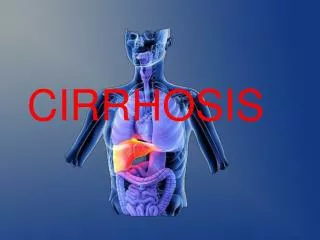
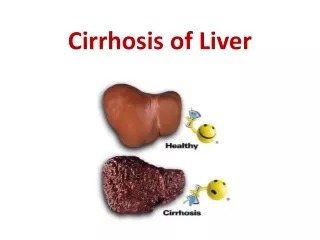
Cirrhosis is a consequence of chronic liver disease characterised by: • replacement of liver tissue by fibrous scar tissue as well as regenerative nodules (lumps that occur as a result of a process in which damaged tissue is regenerated). • leading to progressive loss of liver function.
Microscopically, cirrhosis is characterized by regeneration nodules, surrounded by fibrous septa. In these nodules, regenerating hepatocytes are disorderly disposed. Portal tracts, central veins and the radial pattern of hepatocytes are absent. Fibrous septa are prominent and may present inflammatory infiltrate (lymphocytes, macrophages). If it is a secondary biliary cirrhosis, biliary ducts are damaged, proliferated or distended - bile stasis. These dilated ducts contain inspissated bile which appear as bile casts or bile thrombi (brown-green, amorphous). Bile retention may be found also in the parenchyma, as the so called "bile lakes.
The pathological hallmark of cirrhosis is: • the development of scar tissue that replaces normal parenchyma, blocking the portal flow of blood through the organ and disturbing normal function. • recent research shows the pivotal role of a stellate cell, a cell type that normally stores vitamin A, in the development of cirrhosis. • damage to the hepatic parenchyma leads to activation of the stellate cell, which becomes contractile (called myofibroblast) and obstructs blood flow in the circulation. • in addition, it secretes TGF-β1, which leads to a fibrotic response and proliferation of connective tissue. • furthermore, it disturbs the balance between matrix metalloproteinases and the naturally occurring inhibitors (TIMP 1 and 2), leading to matrix breakdown and replacement by connective tissue-secreted matrix.
CAUSES OF CIRRHOSIS • Cirrhosis is most commonly caused by: • Alcohol • Hepatitis B and C • Fatty liver disease • cryptogenic, i.e. of unknown cause
The word "cirrhosis" derives from Greek kirrhos, meaning "tawny" (the orange-yellow colour of the diseased liver). While the clinical entity was known before, it was Laennec who gave it the name "cirrhosis" in his 1819 work in which he also describes the stethoscope
CIRRHOSIS STATISTICS • Cirrhosis and chronic liver disease were the 10th leading cause of death for men and the 12th for women in the United States in 2001, killing about 27,000 people each year. • Established cirrhosis has a 10-year mortality of 34-66%, largely dependent on the cause of the cirrhosis. • The risk of death due to all causes is increased twelvefold; if one excludes the direct consequences of the liver disease, there is still a fivefold increased risk of death in all disease categories. • Little is known on modulators of cirrhosis risk, apart from other diseases that cause liver injury (such as the combination of alcoholic liver disease and chronic viral hepatitis, which may act synergistically in leading to cirrhosis). • Studies have recently suggested that coffee consumption may protect against cirrhosis, especially alcoholic cirrhosis.
CIRRHOSIS: CLINICAL PRESENTATION • Spider nevi: vascular lesions consisting of a central arteriole surrounded by many smaller vessels due to an increase in oestradiol. These occur in about 1/3 of cases. • Palmar erythema: exaggerations of normal speckled mottling of the palm, due to altered sex hormone metabolism. • Nail changes: • Muehrcke's nails - paired horizontal bands separated by normal colour due to hypoalbuminaema • Terry’s nails - proximal two thirds of the nail plate appears white with distal one-third red, also due to hypoalbuminemia • Clubbing - angle between the nail plate and proximal nail fold > 180 degrees • Hypertrophic osteoarthropathy:chronic proliferative periostitis of the long bones that can cause considerable pain. • Dupytren’s contracture: thickening and shortening of palmar fascia that leads to flexion deformities of the fingers. Common (33% of patients).
Gynaecomastia • Hypogonadism • Hepatomegaly • Splenomegaly • Oesophageal varices
Ascites: accumulation of fluid in the peritoneal cavity giving rise to flank dullness (needs about 1500 mL to detect flank dullness). It may be associated with hydrocoele and penile flomation (swelling of the penile shaft) in men. • Caput medusae:in portal hypertension, the umbilical vein may open. Blood from the portal venous system may be shunted through the periumbilical veins into the umbilical vein and ultimately to the abdominal wall veins, manifesting as caput medusa. • Cruveilhier-Baumgarten murmur: venous hum heard in epigastric region (on examination by stethoscope) due to collateral connections between portal system and the remnant of the umbilical vein in portal hypertension. • Foetor hepaticus: musty odour in breath due to increased dimethyl sulphide.
Jaundice: yellow discolouring of the skin, eye, and mucus membranes due to increased bilirubin (at least 2-3 mg/dL or 30 mmol/L). Urine may also appear dark. • Asterixis: bilateral asynchronous flapping of outstretched, dorsiflexed hands seen in patients with hepatic encephalopathy. • Other: weakness, fatigue, anorexia, weight loss.
As the disease progresses, complications may develop. • In some people, these may be the first signs of the disease. • Bruising and bleeding due to decreased production of coagulation factors. • Jaundice due to decreased processing of bilirubin. • Itching (pruritus) due to bile products deposited in the skin. • Hepatic encephalopathy - the liver does not clear ammonia and related nitrogenous substances from the blood, which are carried to the brain, affecting cerebral functioning: • neglect of personal appearance, • unresponsiveness, • forgetfulness, • trouble concentrating, • changes in sleep habits.
Sensitivity to medication due to decreased metabolism of the active compounds. • Hepatocellular carcinoma is primary liver cancer, a frequent complication of cirrhosis. • Portal hypertension - blood normally carried from the intestines and spleen through the hepatic portal vein flows more slowly and the pressure increases; this leads to the following complications: • - Ascites - fluid leaks through the vasculature into the abdominal cavity. • - Esophageal varices - collateral portal blood flow through vessels in the stomach and esophagus. These blood vessels may become enlarged and are more likely to burst.
Multi-system effects: • Cirrhosis can cause immune system dysfunction, leading to infection. • Fluid in the abdomen (ascites) may become infected with bacteria normally present in the intestines (spontaneous bacterial peritonitis). • Hepatorenal syndrome - insufficient blood supply to the kidneys, causing acute renal failure. This complication has a very high mortality (over 50%). • Hepatopulmonary syndrome - blood bypassing the normal lung circulation (shunting), leading to cyanosis and dyspnoea (shortness of breath), characteristically worse on sitting up. • Portopulmonary hypertension - increased blood pressure over the lungs as a consequence of portal hypertension.[6]
Alcoholic liver disease (ALD); • - Alcoholic cirrhosis develops in 15% of individuals who drink heavily for more than a decade. • There is great variability in the amount of alcohol needed to cause cirrhosis (as little as 3-4 drinks a day in some men and 2-3 in some women. • Patients may also have concurrent alcoholic hepatitis with fever, hepatomegaly, jaundice, and anorexia. • AST and ALT are both elevated but less than 300 IU/L with a AST:ALT ratio > 2.0, a value rarely seen in other liver diseases. • Liver biopsy may show hepatocyte necrosis, Mallory bodies, neutrophilic infiltration with perivenular inflammation.
Chronic hepatitis C. • - low grade damage to the liver that over several decades can lead to cirrhosis. • Chronic hepatitis B. • The hepatitis B virus is probably the most common cause of cirrhosis worldwide, especially South-East Asia, but it is less common in the United States and the Western world. • Hepatitis B causes liver inflammation and injury that over several decades can lead to cirrhosis. • Hepatitis D is dependent on the presence of hepatitis B, but accelerates cirrhosis in co-infection.
Non-alcoholic steatohepatitis (NASH): • In NASH, fat builds up in the liver and eventually causes scar tissue. • This type of hepatitis appears to be associated with diabetes, protein malnutrition, obesity, coronary artery disease, and treatment with corticosteroid medications. • This disorder is similar to that of alcoholic liver disease but patient does not have an alcohol history. • Biopsy is needed for diagnosis.
Primary biliary cirrhosis: • May be asymptomatic or complain of fatigue, pruritus, and non-jaundice skin hyperpigmentation with hepatomegaly. • Prominent alkaline phosphatase elevation as well as elevations in cholesterol and bilirubin. • Gold standard diagnosis is antimitochondrial antibodies with liver biopsy as confirmation if showing florid bile duct lesions. It is more common in women. • Primary sclerosing cholangitis: • PSC is a progressive cholestatic disorder presenting with pruritus, steatorrhea, fat soluble vitamin deficiencies, and metabolic bone disease. • - There is a strong association with inflammatory bowel disease (IBD), especially ulcerative colitis. • Diagnosis is best with contrast cholangiography showing diffuse, multifocal strictures and focal dilation of bile ducts, leading to a beaded appearance. • Non-specific serum immunoglobulins may also be elevated.
Autoimmune hepatitis: • This disease is caused by the immunologic damage to the liver causing inflammation and eventually scarring and cirrhosis. • Findings include elevations in serum globulins, especially gamma globulins. • Therapy with prednisone +/- azathioprine is beneficial. • Cirrhosis due to autoimmune hepatitis still has 10-year survival of 90%+. • There is no specific tool to diagnose autoimmune but it can be beneficial to initiate a trial of corticosteroids.
Hereditary haemochromatosis: • Usually presents with family history of cirrhosis, skin hyperpigmentation, diabetes mellitus, pseudogout, and/or cardiomyopathy, all due to signs of iron overload. • Laboratory investigation will show fasting transferrin saturation of > 60% and ferritin > 300 ng/mL. • Genetic testing may be used to identify HFE mutations. If these are present, biopsy may not need to be performed. • Treatment is with phlebotomy to lower total body iron levels.
Wilson's disease: • Autosomal recessive disorder characterized by low serum ceruloplasmin and increased hepatic copper content on liver biopsy. May also have Kayser-Fleischer rings in the cornea and altered mental status. • Alpha 1-antitrypsin deficiency (AAT): • Autosomal recessive disorder. Patients may also have COPD, especially if they have a history of tobacco smoking. Serum AAT levels are low. Recombinant AAT is used to prevent lung disease due to AAT deficiency. • Cardiac cirrhosis: • Due to chronic right sided heart failure which leads to liver congestion. • Galactosemia • Glycogen storage disease type IV • Cystic fibrosis • Drugs or toxins • Certain parasitic infections (such as schistosomiasis)
DIAGNOSIS • The gold standard for diagnosis of cirrhosis is a liver biopsy, through a per-cutaneous, trans-jugular, laparoscopic, or fine-needle approach. • Histologically cirrhosis can be classified as micronodular, macronodular, or mixed, but this classification has been abandoned since it is non-specific to the aetiology, it may change as the disease progresses, and serological markers are much more specific. • However, a biopsy is not necessary if the clinical, laboratory, and radiologic data suggests cirrhosis. • Furthermore, there is a small but significant risk to liver biopsy, and cirrhosis itself predisposes for complications due to liver biopsy.
LABORATORY FINDINGS [1]: • Aminotransferases - AST and ALT are moderately elevated, with AST > ALT. However, normal aminotransferases do not preclude cirrhosis. • Alkaline phosphatase - usually slightly elevated. • GGT – correlates with AP levels. Typically much higher in chronic liver disease from alcohol. • Bilirubin - may elevate as cirrhosis progresses. • Albumin - levels fall as the synthetic function of the liver declines with worsening cirrhosis since albumin is exclusively synthesized in the liver • Prothrombin time - increases since the liver synthesizes clotting factors.
LABORATORY FINDINGS [2]: • Globulins- increased due to shunting of bacterial antigens away from the liver to lymphoid tissue. • Serum sodium - hyponatraemia due to inability to excrete free water resulting from high levels of ADH and aldosterone. • Thrombocytopenia - due to both congestive splenomegaly as well as decreased thrombopoietin from the liver. However, this rarely results in platelet count < 50,000/mL. • Leukopenia and neutropenia - due to splenomegaly with splenic margination. • Coagulation defects - the liver produces most of the coagulation factors and thus coagulopathy correlates with worsening liver disease.
Other laboratory studies performed in newly diagnosed cirrhosis may include: • Serology for hepatitis viruses, autoantibodies (ANA, anti-smooth muscle, anti-mitochondria, anti-LKM) • Ferritin and transferrin saturation (markers of iron overload), copper and ceruloplasmin (markers of copper overload) • Immunoglobulin levels (IgG, IgM, IgA) - these are non-specific but may assist in distinguishing various causes • Cholesterol and glucose • Alpha 1-antitrypsin
RADIOLOGICAL INVESTIGATION [1] Ultrasound is routinely used in the evaluation of cirrhosis, where it may show a small and nodular liver in advanced cirrhosis along with increased echogenicity with irregular appearing areas. Ultrasound may also screen for hepatocellular carcinoma, portal hypertension and Budd-Chiari syndrome (by assessing flow in the hepatic vein).
RADIOLOGICAL INVESTIGATION [2] A new type of device, the FibroScan (transient elastography), uses elastic waves to determine liver stiffness which theoretically can be converted into a liver score based on the METAVIR scale. The FibroScan produces an ultrasound image of the liver (from 20-80mm) along with a pressure reading (in kPa.) The test is much faster than a biopsy (usually last 2.5-5 minutes) and is completely painless. It shows reasonable correlation with the severity of cirrhosis
Other tests performed in particular circumstances include abdominal CT and liver/bile duct MRI (MRCP).
Endoscopy (endoscopic examination of the esophagus, stomach and duodenum) is performed in patients with established cirrhosis to exclude the possibility of esophageal varices. If these are found, prophylactic local therapy may be applied (sclerotherapy or banding) and beta blocker treatment may be commenced. Rarely diseases of the bile ducts, such as primary sclerosing cholangitis, can be causes of cirrhosis. Imaging of the bile ducts, such as ERCP or MRCP (MRI of biliary tract and pancreas) can show abnormalities in these patients, and may aid in the diagnosis.
CLINICAL ASSESSMENT OF SEVERITY • The severity of cirrhosis is commonly classified with the Child-Pugh score. • This score uses bilirubin, albumin, INR, presence and severity of ascites and encephalopathy to classify patients in class A, B or C: • class A has a favourable prognosis, • class C is at high risk of death. • It was devised in 1964 by Child and Turcotte and modified in 1973 by Pugh et al.
More modern scores, used in the allocation of liver transplants but also in other contexts, are: • the Model for End-Stage Liver Disease (MELD) score and its pediatric counterpart, • the Paediatric End-Stage Liver Disease (PELD) score. • The hepatic venous pressure gradient, i.e. the difference in venous pressure between afferent and efferent blood to the liver, also determines severity of cirrhosis, although hard to measure. • A value of 16 mm or more means a greatly increased risk of dying.
Treatment Generally, liver damage from cirrhosis cannot be reversed, but treatment could stop or delay further progression and reduce complications. A healthy diet is encouraged, as cirrhosis may be an energy-consuming process. Close follow-up is often necessary. Antibiotics will be prescribed for infections, and various medications can help with itching. Laxatives, such as lactulose, decrease risk of constipation; their role in preventing encephalopathy is limited.