Download
1 / 20
220 likes | 397 Vues
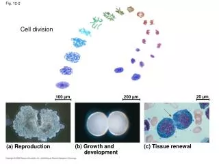
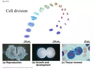
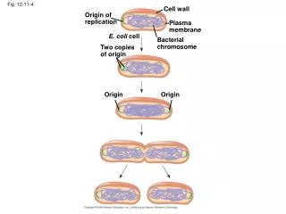
Cell wall. Origin of replication. Plasma membrane. E. coli cell. Bacterial chromosome. Two copies of origin. Fig. 12-11-4. Origin. Origin. 20 µm. 100 µm. 200 µm. Fig. 12-2. (a) Reproduction. (b) Growth and development. (c) Tissue renewal. INTERPHASE. S

E N D
Cell wall Origin of replication Plasma membrane E. coli cell Bacterial chromosome Two copies of origin Fig. 12-11-4 Origin Origin
20 µm 100 µm 200 µm Fig. 12-2 (a) Reproduction (b) Growth and development (c) Tissue renewal
INTERPHASE S (DNA synthesis) G1 Fig. 12-5 Cytokinesis G2 Mitosis MITOTIC (M) PHASE
0.5 µm Chromosomes DNA molecules Chromo- some arm Chromosome duplication (including DNA synthesis) Fig. 12-4 Centromere Sister chromatids Separation of sister chromatids Centromere Sister chromatids
Fig. 12-6 Metaphase Anaphase Telophase and Cytokinesis G2 of Interphase Prophase Prometaphase Centrosomes (with centriole pairs) Early mitotic spindle Centromere Chromatin (duplicated) Fragments of nuclear envelope Nonkinetochore microtubules Aster Cleavage furrow Metaphase plate Nucleolus forming Daughter chromosomes Nuclear envelope forming Centrosome at one spindle pole Spindle Nuclear envelope Kinetochore Chromosome, consisting of two sister chromatids Kinetochore microtubule Plasma membrane Nucleolus
Fig. 12-6a Prometaphase G2 of Interphase Prophase
G2 of Interphase Prophase Prometaphase Chromatin (duplicated) Centrosomes (with centriole pairs) Early mitotic spindle Fragments of nuclear envelope Centromere Aster Nonkinetochore microtubules Fig. 12-6b Kinetochore Nuclear envelope Plasma membrane Chromosome, consisting of two sister chromatids Kinetochore microtubule Nucleolus
Fig. 12-6c Metaphase Anaphase Telophase and Cytokinesis
Telophase and Cytokinesis Metaphase Anaphase Nucleolus forming Metaphase plate Cleavage furrow Fig. 12-6d Daughter chromosomes Nuclear envelope forming Centrosome at one spindle pole Spindle
Aster Centrosome Sister chromatids Microtubules Chromosomes Metaphase plate Fig. 12-7 Kineto- chores Centrosome 1 µm Overlapping nonkinetochore microtubules Kinetochore microtubules 0.5 µm
Telophase and Cytokinesis Metaphase Anaphase Nucleolus forming Metaphase plate Cleavage furrow Fig. 12-6d Daughter chromosomes Nuclear envelope forming Centrosome at one spindle pole Spindle
Fig. 12-9 Vesicles forming cell plate Wall of parent cell 1 µm 100 µm Cleavage furrow Cell plate New cell wall Daughter cells Contractile ring of microfilaments Daughter cells (a) Cleavage of an animal cell (SEM) (b) Cell plate formation in a plant cell (TEM)
Fig. 12-9a 100 µm Cleavage furrow Daughter cells Contractile ring of microfilaments (a) Cleavage of an animal cell (SEM)
Fig. 12-9b Vesicles forming cell plate Wall of parent cell 1 µm Cell plate New cell wall Daughter cells (b) Cell plate formation in a plant cell (TEM)
Nucleus Chromatin condensing 10 µm Fig. 12-10 Chromosomes Cell plate Nucleolus 2 4 1 Prophase Prometaphase 3 Metaphase Anaphase Telophase 5
G1 checkpoint Fig. 12-14 Control system S G1 G2 M M checkpoint G2 checkpoint
G0 G1 checkpoint Fig. 12-15 G1 G1 (b) Cell does not receive a go-ahead signal Cell receives a go-ahead signal
M M S G1 G1 M G1 S G2 G2 MPF activity Cyclin concentration Fig. 12-17 Time (a) Fluctuation of MPF activity and cyclin concentration during the cell cycle S G1 Cdk Cyclin accumulation M Degraded cyclin G2 G2 Cdk checkpoint Cyclin is degraded Cyclin MPF (b) Molecular mechanisms that help regulate the cell cycle
Anchorage dependence Fig. 12-19 Density-dependent inhibition Density-dependent inhibition 25 µm 25 µm (a) Normal mammalian cells (b) Cancer cells
Fig. 12-20 Lymph vessel Tumor Blood vessel Cancer cell Glandular tissue Metastatic tumor Cancer cells invade neigh- boring tissue. A tumor grows from a single cancer cell. Cancer cells spread to other parts of the body. Cancer cells may survive and establish a new tumor in another part of the body. 4 2 1 3