The eye
The eye. The eye in situ Section through the eye Focusing an image on the retina Eye defects. Exit. Face view of the eye. eyebrow. Tear gland. Upper eyelid and lashes. pupil. iris. Lower eyelid. Duct draining tear to nose. Exit. Left eye in side view. Rectus muscle. Tear gland.

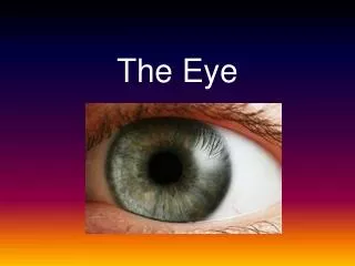
The eye
E N D
Presentation Transcript
The eye • The eye in situ • Section through the eye • Focusing an image on the retina • Eye defects Exit
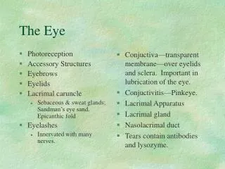
Face view of the eye eyebrow Tear gland Upper eyelid and lashes pupil iris Lower eyelid Duct draining tear to nose Exit
Left eye in side view Rectus muscle Tear gland eyelid Oblique muscle Muscle attachment eyeball orbit Optic nerve Exit
Fovea / yellow spot Suspensory ligament Ciliary muscle lens Aqueous humour pupil Optic nerve iris Vitreous humour cornea conjunctiva sclera choroid Blind spot retina Horizontal section of the eye Exit
Change of Pupil Size • pupil is the opening surrounded by iris allowing light to pass through • pupil limits amount of light entering the eye • two kinds of iris muscle control size of pupil:- circular iris muscle: makes pupil constrict as they contract - radial iris muscle: pupil dilates as they contract
radial muscle relax reduce amount of light pass through pupil size decrease Under Bright Light circular muscle contract
circular muscle relaxes increase amount of light pass through pupil size increases Under Dim Light radial muscle contracts
Radial muscle relaxed Radialmuscle contracted Pupil dilated Pupil constricted Circular muscle contracted Circular muscle relaxed 1.Circular muscle of iris contract 1. Circular muscle of iris relax 2.Radial muscle of iris relax 2.Radial muscle of iris contract 3.Pupil constricts 3. Pupil dilates 4.Less light enters the eye 4.More light enters the eye 5.Depth of focus is increased 5. Depth of focus is decreased In bright light In dim light Exit
Change of pupil size Click for video
shorter focal length longer focal length Accommodation • lens more convex • lens less convex • accommodation is the process of the eye to focus an image on retina i.e. adjustthickness/convexity of lens for viewing near and distant objects . • helped by the action of ciliary muscles
light rays refracted more & images of near objects fall on retina ring of ciliary body moves inwards and becomes smaller in diameter lens becomes thicker & more convex tension of suspensory ligaments reduces Focusing Near Objects ciliary muscles contract
circular ciliary muscle contracts decrease in circumference tensionof suspensory ligament decreases lens becomes more convex
Exit 4. Suspensory ligaments taut 3. Ciliary muscle relaxed 2. Cornea refracts light ray 1. Parallel rays of light from a distant object 5. Lens pulled flat (less convex) 6. Light refracted less 7. Image focused onto retina Focusing on distant objects
image of distant objects fall on retina ring of ciliary body moves outwards and becomes wider in diameter lens becomes thinner/less convex tension of suspensory ligaments increases Focusing Distant Objects ciliary muscles relax
circular ciliary muscle relaxes increase in circumference tension of suspensory ligament increases lens becomes less convex
4. Suspensory ligaments relaxed 3. Ciliary muscle contracted 2. Cornea refracts light ray 1. Diverging rays of light from a near object 5. Lens thickens (more convex) 6. Light refracted more 7. Image focused onto retina Focusing on near objects Exit
Normal eye retina Near object, lens more convex Distant object, lens less convex Exit
Common Eye Defects 1. Short-sightedness 2. Long-sightedness 3. Colour-blindness
Short sightedness Defect Correction Eyeball too long Image of far object fall in front of retina Wear concave lens Concave lens diverges light rays, and image is focused on retina Exit
Long sightedness / far sightedness Defect Correction Eyeball too short Image of near object fall behind the retina Wear convex lens Convex lens converges light rays, and image is focused on retina Exit
Colour Blindness • Cone cells are responsible for colour vision • There are three kinds of cone cells responsible for detecting red, green and blue lights • relative number of cones present & stimulated determines the colour perceived • colour blindness is a hereditary defect and cannot be corrected by wearing glasses
Can You See? Ans: 5 Ans: 8 Ans: Nothing Ans: A line