
Comprehensive Eye Assessment Guide for Nurses
This guide covers pertinent eye history questions, physical assessments, differentiation between normal/abnormal findings, and identifying health problems via nursing diagnoses. Includes structures/functions, interactions with other systems, developmental variations, and a detailed case study. Also provides insight on symptoms signaling eye problems, important history findings, and instructions for visual acuity tests and external eye structure inspections.

Comprehensive Eye Assessment Guide for Nurses
E N D
Presentation Transcript
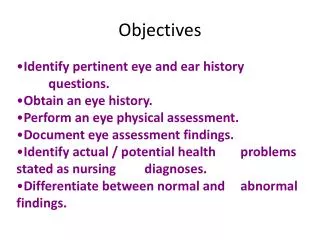
Objectives • Identify pertinent eye and ear history questions. • Obtain an eye history. • Perform an eye physical assessment. • Document eye assessment findings. • Identify actual / potential health problems stated as nursing diagnoses. • Differentiate between normal and abnormal findings.
Structure & Functions External Structures Eyelids and lashes: Protect the eyes Lacrimal glands and ducts: Produce tears Conjunctiva: Provide lubrication
Sclera: Gives shape and structure to eye Iris: Controls amount of light entering eye; provides eye color Extraocular muscles: Control eye movement
Cornea: Transparent, avascular outer layer of the eyeball Anterior chamber: Filled with aqueous humor Pupil: The aperture of the iris
Structure & Functions Internal Structures Optic disc and physiological cup: Area where the optic nerve and the blood vessels enter the eye Retinal blood vessels: Blood supply to eye
Retina: Inner layer; receives light waves that are sent to brain and converted into visible perceptions Macula: Avascular, darker area of central vision
Relationship to Other Systems Integumentary *Skin covers the lids *Lashes protect the eyes Respiratory *Color changes seen in conjunctiva Digestive *Jaundice seen in sclera *Sight of food can stimulate saliva production
Relationship Con’t Cardiovascular *Xanthelasma (fat deposits) can be seen on the eyelids *Can observe blood vessels directly (opthalmoscope) *Observed changes can reveal HTN, Diabetes, Glaucoma, Intracranial Pressure, some CNS disorders Musculoskeletal *Movement of eyeball & lid; Iris adjusts Pupil Neurological CN II responsible for vision CN III, IV, VI responsible for movement of eye CN V conveys sensory data from cornea CN VII closes eyelids
Relationship Con’t • Reproductive Pleasure selection • Urinary Periorbital edema with renal disease Immune Conjunctivitis: Environmental Toxins, Allergies, Infection • Endocrine Thyroid, Diabetes
DevelopmentalVariations What developmental variations of the eyes might be seen with: • Infants: shape/oval, spacing, vision 20/200 • How is vision determined in Infants? pupillary, response, blink response, ability to follow objects
Developmental (Con’t.) • Preschool: Snellen E chart: 3yr old 20/40 4yr old vision 20/30, 5-6yr old normal • School Age: tests: color blindness (pg. 341), regular Snellen • Older adults: All around deterioration
Case Study • Laura 55- year-old secretary, widow • History of HTN, DM • Complains of blurred vision, worsened over past several months • Medications include Lasix, Captopril and Glucatrol
Vision loss Double vision Eye tearing Eye drainage Eye pain Blurred Vision Dry Eyes Eye Appearance Changes What symptoms would signal a problem with the eyes?
Pertinent History Findings • Secretary, 4 hours computer work/day • Blurred vision: constant, and worse with fatigue • Difficult to read, reading glasses not helpful • HTN, DM • Lasix, Captopril, and Glucatrol • + Family history of CV disease, DM, MS • Last eye exam 5 years ago • Widow: son and friends as supports
Physical Assessment • Anatomical Landmarks: visual fields (superior, inferior, nasal, temporal) • Approach: inspection, palpation, ophthalmoscopy • Position: sitting • Tools: visual acuity charts (Snellen), penlight, ophthalmoscope, cotton ball, cotton swab, printed material, pg 264 • General survey and head-to-toe scan
Visual Acuity • Far vision: Snellen eye chart • Near vision: read newsprint 13 to 15” from eyes • Color vision: identify color bars on Snellen or use color plates • Peripheral vision: come in from the periphery in all fields and note field cuts
Inspection of External Structures • Lids and lashes: color, lesions, edema, symmetry, position, crusting and distribution of lashes, infestation • Lacrimal glands and ducts: color, edema, excessive tearing or drainage • Conjunctiva: color, moisture, lesions, and foreign bodies • Sclera: color, moisture, lesions, or tears Should be smooth and white
Inspection of External Structures, Con’t • Cornea: clarity--if cloudy could be a Vit A deficiency, abrasions/ulcers--rough, corneal reflex (cotton ball or puff of air) • Anterior chamber: clarity, is there a bulging iris, blood or pus? • Iris: color, size, shape, and symmetry • Pupils: size, shape, reaction to light
Palpation of External Structures • Eye ball: consistency and tenderness • Lacrimal glands and ducts: tenderness and excessive tearing
Ophthalmoscopy • Red reflex: presence, opacities • Optic disc and physiologic cup: color, size, shape, borders, cup-disc ratio • Retinal vessels: size ratio of arteries and veins, color, arteriole light reflex, crossings • Retina: color, texture, exudates, lesions, hemorrhages, and aneurysms • Macula and fovea: color, size, location, lesions
Bits and Pieces: If mydriatics are used: Instruct to wear sunglasses to protect eyes If examining undilated pupils with scope: use smallest white light aperture in darkened room If using eye chart: 1st test without corrective lenses, then with them (reduces likelihood that the measurement without will be influenced) Glazed eyes: febrile state Asymmetry of lids: CN III damage, CVA Eye exams (American Academy of Ophthalmology): Preschool screening (strabismus can lead to blindness) Every 3-5 yrs for African descent ages 20-39 Every 2-4 yrs for anyone ages 40-64 Every 1-2 yrs starting at age 65 Every 1 yr for DM
Tips for Practice with an Ophthalmoscope • When examining undilated eyes, use the smallest white light aperture. • Start with small round light and lens set “0” • At 1 ft away and 15 degree angle: pupil • When obtain red reflex, move forward • Use R eye to examine R eye & L to exam L • As you move forward place your hand on their head to orient yourself • Adjust the lens focus as needed with a free finger


