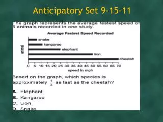
Anticipatory Set 9-15-11
580 likes | 896 Vues
Anticipatory Set 9-15-11. Anticipatory Set 9-21-11 L2&3. Cellular Transport. Selective Permeability. Selective permeability- the cell membrane’s ability to allow some substances to enter/ exit but not all. Two processes that allow substances to enter/exit:

Anticipatory Set 9-15-11
E N D
Presentation Transcript
Selective Permeability • Selective permeability- the cell membrane’s ability to allow some substances to enter/ exit but not all. • Two processes that allow substances to enter/exit: • Passive transport – energy from kinetic energy and concentration gradient. • Active transport-ATP
Diffusion • Process depends on concentration gradient. • Particles will never stop moving, but when equilibrium is reached there will be no net change in their concentration. • Movement of particles is from [high] to [low] concentration. • Dependent on four factors: diameter, temperature, electrical charge (when applicable), and the concentration gradient. • Majority of materials enter cell through diffusion…energy conservation for other processes.
Cell Membrane & Diffusion • diffusion animation
Osmosis • Diffusion of water. • Water always travels from hypotonic to hypertonic • Solute always travels in opposite direction of water. • Osmosis Animation
Isotonic Solutions • Isotonicsolutions-the same amount of solute exists inside and outside the cell. • Water moves in and out at the same rate.
Hypertonic Solutions • Hypertonic solutions- have more solutes in solution than inside the cell. • Water moves out of the cell to achieve equilibrium.
Hypotonic Solutions • Hypotonic solutions- have less solutes in them than inside the cell. • Water will enter the cell to try and achieve equilibrium. • Cells may lyse if too much water enters. • Plants combat this risk with their cell wall, and turgor pressure results.
Osmoregulation-water control • Turgid- enough water, plant cell rigid • Flaccid- lacking water, plant cell limp • Plasmolysis- cell membrane is ripped from cell wall
Isotonic solution Hypertonic solution Hypotonic solution Animal cell H2O H2O H2O H2O . Shriveled Normal Lysed Plant cell H2O H2O H2O H2O Flaccid Plasmolyzed Turgid (normal)
Water Potential- water’s ability to do work when going through the C.M. • Pressure Potential • Positive pressure is the cell being pushed • Negative Pressure- the cell being pulled (eg transpiration) • Solute Potential- based on solute concentration
Positive Pressure Potential Positive Pressure Potential
Negative Pressure Potential Negative Pressure Potential
Solute Potential ΨS = -iCRT -i (ionization constant) C (molar concentration) R (pressure constant) T (temperature in Kelvin)
Turgor pressure is ~100psi, much more than a tire. The pressure is so great that plant cells would detach from one another if not for adhesive molecules known as pectins.
Facilitated Diffusion with Channel Proteins • Facilitated diffusion- within the cell membrane are channel proteins that allow materials to pass into the cell. • Aquaporins- channel proteins that allow water to pass through, in addition to simple diffusion. • In kidneys and plants where water is essential • Channel Protein Animation
FD with Ion Channel Proteins • Channel protein let in ions. • When the protein shape changes, the gate will open. • Ions pass through based on size and charge. • Can be ligand gated or voltage gated. • Voltage gated channels depend on two things: • Concentration gradient of K • Concentration of K (usually higher inside cell) • Membrane potential due to charge imbalance.
Facilitated Diffusion with Carrier Proteins • Carrier proteins transport polar substances like amino acids and sugars. • When the carrier proteins become saturated the rate of diffusion is maxed out. • Animation: How Facilitated Diffusion Works
Figure 5.12 A Carrier Protein Facilitates Diffusion (Part 1)
Filtration • Filtration- pressure driven system that pushes water and nutrients across cell membranes. • This is how urine is produced • Does not require energy.
Active Transport is Directional • Active transport always works against the concentration gradient. Going from a lower to higher concentration. • Requires energy. • Two types: primary and secondary active transport.
Membrane Proteins associated with Active Transport • Cell Pumps: • Uniports move a single substance in one direction. • Symports – move two substances in the same direction. • Antiports - move two substances in opposite directions. One into the cell, and one out of the cell. • e.g. NaK pump • Coupled transporters are those that move two substances. Which of these are coupled?
Primary Active Transport • ATP is hydrolyzed and drives the movement of ions against the concentration gradient. • Sodium potassium pump is an example of 1AT. Because the ions move against the concentration gradient. (Na leaves cell, although more Na outside cell, same with K more in cell, but K still enters) • NaK Pump located in all animal cells; antiport; coupled transporter • NaK Pump Simple Animation • Na K Pump Animation
Figure 5.14 Primary Active Transport: The Sodium–Potassium Pump
Membrane Potential • Membrane Potential aka Voltage Gradient allows the cell to do work. • DNA is negative inside cell(-), NaK pumps extra Na out of the cell (+). • Difference in charge allows molecules to be transported using ATP. • E.g. glucose enters through because of membrane potential • Secondary AT Animation
H Pumps • Most important pump for cell respiration and photosynthesis. • H+ pumped out of cell, and ions can now diffuse in • Pumping H requires little energy, and they help sugars enter the cell by AT
What if the macromolecules are too large, charged, or polar to enter through the membrane? • Is this a good problem or not? • Which organelle is responsible for substance transport?
Endocytosis • Processes that bring substances into the cell such as macromolecules and smaller cells. • Three types of endocytosis: • Phagocytosis • Pinocytosis • Receptor-Mediated Endocytosis
Figure 5.16 Endocytosis and Exocytosis (A) Phagocytosis • Cell eating • Part of cell membrane engulfs particles/cells • Phagosome fuses with a lysosome and digestion occurs
Endocytosis • Phagocytosis- process fairly nonspecific • Only a few cells can do this ex. WBC • Must be able to change shape and form pseudopodia. • WBC will attach to bacteria engulf bacteria with pseudopodia lysosomes with enzymes digest it residual waste is exocytosed. • Pinocytosis- same process just with liquids. Also fairly nonspecific. • WBC and Phagocytosis Animation
Receptor-Mediated Endocytosis • Specific process that utilizes integral membrane proteins to bind to specific molecules in the cell’s environment. • Receptor proteins are substance specific, aka coated pits. Coated with protein , formed by CM depressions. • When a ligand binds to the receptor protein, it invaginates and forms a vesicle. • E.g. cholesterol uptake in mammals rd. 113-114
Figure 5.17 Formation of a Coated Vesicle (Part 2) Receptors will form a new vesicle and be recycled back to plasma membrane.
Exocytosis • Anything that comes in must go out. • Materials are packaged into vesicles, which fuse with the cell membrane via a membrane protein. • The two membranes fuse, contents expelled, and the CM incorporates vesicle membrane.
Endocytosis and Exocytosis Animation Hyper,Hypo,Iso