
Muscle Structure and Contraction Mechanisms
E N D
Presentation Transcript
Muscle XIA Qiang, MD & PhD Department of Physiology Room 518, Block C, Research Building School of Medicine, Zijingang Campus Email: xiaqiang@zju.edu.cn Tel: 88206417 (Undergraduate school), 88208252 (Medical school)
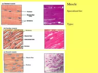
Muscle Types of muscle: • Skeletal muscle • Cardiac muscle • Smooth muscle Striated muscle
Muscle (cont.) • The sliding filament mechanism, in which myosin filaments bind to and move actin filaments, is the basis for shortening of stimulated skeletal, smooth, and cardiac muscles. • In all three types of muscle, myosin and actin interactions are regulated by the availability of calcium ions. • Changes in the membrane potential of muscles are linked to internal changes in calcium release (and contraction).
Muscle (cont.) • Neuronal influences on the contraction of muscles is affected when neural activity causes changes in themembrane potential of muscles. • Smooth muscles operate in a wide variety of involuntaryfunctions such as regulation of blood pressure andmovement of materials in the gut.
Skeletal muscles are attached to the skeleton by tendons. Skeletal muscles typically contain many, many muscle fibers
The sarcomere is composed of: thick filaments called myosin, anchored in place by titin fibers, and thin filaments called actin, anchored to Z-lines .
A cross section through a sarcomere shows that: • each myosin can interact with 6 actin filaments, and • each actin can interact with 3 myosin filaments.
Myosin filament (thick filament) • Myosin
Actin filament (thin filament) • Actin • Tropomyosin • Troponin
Sarcotubular system (1) Transverse Tubule (2) Longitudinal Tubule Sarcoplasmic reticulum
Molecular mechanisms of contraction Sliding-filament mechanism
Contraction (shortening): myosin binds to actin, and slides it, pulling the Z-lines closer together, and reducing the width of the I-bands. Note that filament lengths have not changed.
Contraction: myosin’s cross-bridges bind to actin; the crossbridges then flex to slide actin.
Click here to play the Sarcomere Shortening Flash Animation
The thick filament called myosin is actually a polymer of myosin molecules, each of which has a flexible cross-bridge that binds ATP and actin.
4. Partial hydrolysis of the bound ATP energizes or “re-cocks” the bridge. 2. The full hydrolysis and departure of ADP + Picauses the flexing of the bound cross-bridge. 3. Binding of a “new” ATP to the cross-bridge uncouples the bridge. The myosin-binding site on actin becomes available, so the energized cross-bridge binds. 1. The cross-bridge cycle requires ATP
The myosin-binding site on actin becomes available, so the energized cross-bridge binds. 1.
2. The full hydrolysis and departure of ADP + Picauses the flexing of the bound cross-bridge.
3. Binding of a “new” ATP to the cross-bridge uncouples the bridge.
4. Partial hydrolysis of the bound ATP energizes or “re-cocks” the bridge.
4. Partial hydrolysis of the bound ATP energizes or “re-cocks” the bridge. 2. The full hydrolysis and departure of ADP + Picauses the flexing of the bound cross-bridge. 3. Binding of a “new” ATP to the cross-bridge uncouples the bridge. The myosin-binding site on actin becomes available, so the energized cross-bridge binds. 1. The cross-bridge cycle requires ATP
Click here to play the Cross-bridge cycle Flash Animation
Roles of troponin, tropomyosin, and calcium in contraction In relaxed skeletal muscle, tropomyosin blocks the cross-bridge binding site on actin. Contraction occurs when calcium ions bind to troponin; this complex then pulls tropomyosin away from the cross-bridge binding site.
Excitation-contraction coupling • Transmission of action potential (AP) along T tubules • Calcium release caused by T tubule AP • Contraction initiated by calcium ions
The latent period between excitation and development of tension in a skeletal muscle includes the time needed to release Ca++ from sarcoplasmic reticulum, move tropomyosin, and cycle the cross-bridges.
The transverse tubules bring action potentials into the interior of the skeletal muscle fibers, so that the wave of depolarization passes close to the sarcoplasmic reticulum, stimulating the release of calcium ions. The extensive meshwork of sarcoplasmic reticulum assures that when it releases calcium ions they can readily diffuse to all of the troponin sites.
Passage of an action potential along the transverse tubule opens nearby voltage-gated calcium channels, the “ryanodine receptor,” located on the sarcoplasmic reticulum, and calcium ions released into the cytosol bind to troponin. The calcium-troponin complex “pulls” tropomyosin off the myosin-binding site of actin, thus allowing the binding of the cross-bridge, followed by its flexing to slide the actin filament. Dihydropyridine (DHP) receptor
General process of excitation and contraction in skeletal muscle • Neuromuscular transmission • Excitation-contraction coupling • Muscle contraction
A single motor unit consists of a motor neuron and all of the muscle fibers it controls.
The neuromuscular junction is the point of synaptic contact between the axon terminal of a motor neuron and the muscle fiber it controls. Action potentials in the motor neuron cause acetylcholine release into the neuromuscular junction. Muscle contraction follows the delivery of acetylcholine to the muscle fiber.
1. The exocytosis of acetylcholine from the axon terminal occurs when the acetylcholine vesicles merge into the membrane covering the terminal. 2. On the membrane of the muscle fiber, the receptors for acetylcholine respond to its binding by increasing Na+ entry into the fiber, causing a graded depolarization. 3. The graded depolarization typically exceeds threshold for the nearby voltage-gate Na+ and K+ channels, so an action potential occurs on the muscle fiber.
Click here to play the Neuromuscular Junction Flash Animation
Click here to play the Action Potentials and Muscle Contraction Flash Animation
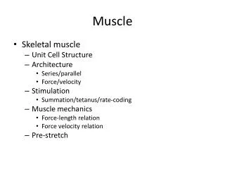
Mechanics of single-fiber contraction • Muscle tension – the force exerted on an object by a contracting muscle • Load – the force exerted on the muscle by an object (usually its weight) • Isometric contraction – a muscle develops tension but does not shorten (or lengthen) (constant length) • Isotonic contraction – the muscle shortens while the load on the muscle remains constant (constant tension)
Twitch contraction • The mechanical response of a single muscle fiber to a single action potential is know as a TWITCH
iso = same tonic = tension metric = length Tension increases rapidly and dissipates slowly Shortening occurs slowly, only after taking up elastic tension; the relaxing muscle quickly returns to its resting length.
All three are isotonic contractions. • Latent period • Velocity of shortening • Duration of the twitch • Distance shortened
Click here to play the Mechanisms of Single Fiber Contraction Flash Animation
Frequency-tension relation Complete dissipation of elastic tension between subsequent stimuli. S3 occurred prior to the complete dissipation of elastic tension from S2. S3 occurred prior tothe dissipation of ANY elastic tension from S2. T e m p o r a l s u m m a t i o n.
Unfused tetanus: partial dissipation of elastic tension between subsequent stimuli. Fused tetanus: no time for dissipation of elastic tension between rapidly recurring stimuli. Frequency-tension relation