The Eye
The Eye. Splawn. Eye. The human eye is 1 inch in diameter. The Protected Eye. The eye is protected by the… Orbital cavity Eyebrows Eyelashes Eyelids. Tears. The eyes are continually bathed by tears secreted out of our lacrimal gland Tears cleanse and moisten. Lacrimal Ducts.

The Eye
E N D
Presentation Transcript
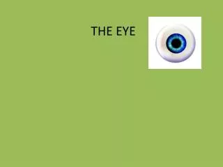
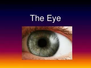
The Eye Splawn
Eye • The human eye is 1 inch in diameter
The Protected Eye • The eye is protected by the… • Orbital cavity • Eyebrows • Eyelashes • Eyelids
Tears • The eyes are continually bathed by tears secreted out of our lacrimal gland • Tears cleanse and moisten
Lacrimal Ducts • Tears flow across the into the lacrimal ducts • The lacrimal ducts empty tears into the nasal cavity
Lubrication from eye lids • Along the border of the eyelid are glands that secrete an oily lubricant for the eyes • An infection here is called a stye
Conjunctiva • A thin membrane that lines the eyelid and covers part of the eye • The conjunctiva secrete mucus which helps lubricate the eye
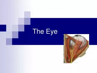
The Wall of the Eye • The wall of the eye is made up of three layers • Sclera • Choroid • Retina
Sclera • The outer layer of the wall of the eye • White of the eye • Tough strong protective layer that helps eye maintain shape • Muscles that move the eye in the socket are attached to the sclera
Extrinsic Muscles • Those muscles attached to the sclera that are responsible for moving the eye within the orbital socket are called extrinsic muscles • 6 different extrinsic muscles the control different eye movements
CLOSE YOUR EYES IF YOU DO NOT WANT TO SEE A REAL EYEBALL WITH REAL EXTRINSIC MUSCLES
Cornea • In the front, center of the sclerotic coat lies a circular clear area called the cornea. • Window of the eye • Transparent to permit passage of light rays • Fed by lymph fluid as transparent due to lack of blood vessels
Cornea (within the sclerotic coat) • Consist of 5 layers of flat cells arranged much like five sheets of glass • Cornea has pain and touch receptors which helps us protect our eyes
Choroid Coat • The middle layer of the wall of the eye • Contains blood vessels that nourish the eye • Darker color to prevent light reflection that would distract from normal vision
Pupil • In the front of the choroid coat there is a dark circular opening that allows for passage of light
Iris • The pupil is surrounded by a colored muscular layer called the iris • Responsible for controlling the diameter and size of the pupil and the amount of light reaching the pupil • Muscles in the iris that control pupil size are called intrinsic muscles
Pupil • Dilated by the contracting intrinsic muscles in dark light or when trying to see an object far away. • Constricted by bright light and close objects
Lens • A crystalline (able to be seen through with clarity) structure located behind the iris and the pupil • Elastic, disc-shaped • Biconvex-curves outward on both sides • Posterior portion more curved that anterior surface • Flattens as we get older
Lens • The lens is held in place by suspensory ligaments from a ciliary body • Ciliary body holds muscles that can change shape of lens • The loose their elasticity with age
Lens • The lens changes shape to allow for near or distant vision • Accommodation
Lens • The lens is situated between the ANTERIOR and POSTERIOR CHAMBERS • The anterior chamber is the space between the cornea and the crystalline lens • The posterior chamber is the space between the iris and the crystalline lens
Lens • The anterior chamber is filled with aqueous humor, a watery fluid that helps maintains pressure in the eye. • The posterior chamber is filled with vitreous humor, jelly-like substance
Retina • Inner-most or third coat of the eye • Between posterior chamber and choroid coat • Does not extend around front portion of eye • It is from this light-sensitive layer that light rays from an object form an image • After the image is focused on the retina, it travels to the optic nerve
Lens • The lens will chan
Positioning of the eye • The location of our eyes in front of the head helps us see stereoscopically • In three dimensions, 3-D • Length • Width • depth