Download
1 / 55
1.58k likes | 4.79k Vues
Serum Protein Electrophoresis. Dr. Nitin Inamdar Department of Biochemistry Tata Memorial Center, Parel, Mumbai. Electrophoresis. Phoresis: Separation or migration or movement Electro: Under influence of electric field. Mixture of proteins Amino acids: Amino group (NH2)

E N D
Serum Protein Electrophoresis Dr. Nitin Inamdar Department of Biochemistry Tata Memorial Center, Parel, Mumbai.
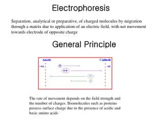
Electrophoresis • Phoresis: Separation or migration or movement • Electro: Under influence of electric field
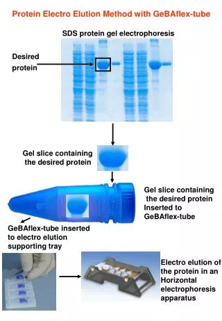
Mixture of proteins • Amino acids: • Amino group (NH2) • Carboxyl group (COOH) • Two amino acids join each other with polypeptide bond to form polypeptide chain & many polypeptide chains make a protein. • Ph 8.6/0.075M Barbitone buffer • 1%Agarose • Constant current 5mAM/Slide Serum Electrophoresis
Electrophoresis separates proteins based on their physical properties. • Serum is placed on a specific medium, and a charge is applied. • The net charge (positive or negative) and the size and shape of the subsets of these proteins are used in interpreting the results.
Serum Electrophoresis Paper electrophoresis Starch gel electrophoresis Agarosre gel electrophoresis Cellulose acetate gel electrophoresis Capillary gel electrophoresis
Serum protein electrophoresis on agarose gel is a type of horizontal gel electrophoresis
Equipment used for the gel electrophoresis power supply (direct current) containers for staining and destaining gel electrophoresis chamber applicator
Various electrophoresis • Serum/protein electrophoresis: • plasma cell dyscrasia, nephrotic syndrome, etc. • Hemoglobin electrophoresis: • thalassemia, hemoglobinopathies like sickle cell anemia, HbC, HbD, etc. • Urine electrophoresis: • BJP in plasma cell dyscrasia
Indication of serum electrophoresis • Diagnostic: • Diagnosis of plasma cell dyscrasia • Diagnosis of Waldenstrom’ macroglbulinemia • Monitoring of disease: • Monitoring of plasma cell dyscrasia
Method of detection • Serum protein electrophoresis • Quantitative immunoglobulins (nephelometry) • Immunofixation, immunoelectrophoresis and immunodiffusion • Urine studies • light chain
Albumin • Globulins: • α1, • α2, • β1, • β2, • gamma • A/G Ratio: 2.0
Anode +ve Cathode -ve Normal serum electrophoresis pH=8.6, 1% agarose gel, sodium barbitone buffer
Anode +ve Cathode -ve Abnormal serum electrophoresis showing M band
WHO Classification of Plasma Cell Neoplasms • Plasma cell myeloma (MM) • Plasma cell myeloma variants Non secretory myeloma Indolent Myeloma Smoldering Myeloma Plasma cell leukemia • Plasmacytomas Solitary plasmacytoma of bone Extramedullary plasmacytoma • Immunoglobulin deposition diseases Primary Amyloidosis Systemic light and heavy chain deposition disease • Osteosclerotic myeloma (POEMS syndrome) • Heavy chain disease Gamma /Mu / Alpha
Diagnostic criteria for plasma cell dyscrasia Symptomatic plasma cell myeloma: BM clonal plasma cells or Plasmacytoma Marrow plasmacytosis (>30%) M-component in serum or urine Serum: Ig G >3 g/dL, Ig A >2.5 g/dL or Urine: >/= 1g/ day of λ or κ [BJP] Asymptomatic plasma cell myeloma: M band in serum at myeloma levels > 3 gm/dL and /or 10% or more clonal plasma cell in BM No related organ or tissue impairment (end organ damage or bony lesion) or myeloma-related symptoms MUGS: M-Component, present but less than 3 gm/dL Marrow Plasmacytosis < 10% No Lytic bone lesions or related organ impairment etc
Immunoglobin (Ig) structure and types Malignant Plasma Cell (Clonal Proliferation) produces one type of Ig either A,M, G, D or E with kappa or lambda light chains in excess amount.
Nephelometry • Antigen-antibody complexes, when formed at a high rate, will precipitate out of a solution resulting in a turbid or cloudy appearance. • Nephelometry: is indirect measurement of light scattered by the antigen-antibody complexes.
Immunofixation electrophoresis • After separation of serum proteins by electrophoresis separated serum in different lanes is incubated with monospecific anti sera against IgA, M G and κλ