High Resolution Protein Electrophoresis
830 likes | 1.6k Vues
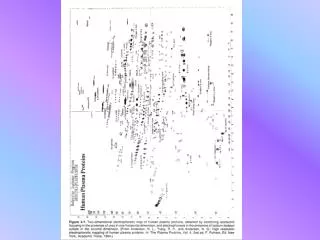
High Resolution Protein Electrophoresis. A Clinical Overview with Case Studies. By Lawrence M. Killingsworth, Ph.D. Only ~ 200 of the vast array of human proteins have been characterized.

High Resolution Protein Electrophoresis
E N D
Presentation Transcript
High Resolution Protein Electrophoresis A Clinical Overview with Case Studies By Lawrence M. Killingsworth, Ph.D.
Only ~200 of the vast array of human proteins have been characterized. Clinical knowledge is limited to 25 – 30 relatively high concentration components of plasma, CSF, urine and other fluids. Of these, 15 or so can be visualized by high resolution agarose electrophoresis. Human Proteins
Normal Control Pre-albumin 1-Acid Glycoprotein -Lipoprotein 1-Antichymotrypsin Haptoglobin C-3 IgA IgG Albumin 1-Antitrypsin 2-Macroglobulin Transferrin -Lipoprotein Fibrinogen IgM
Uncontrolled proliferation of a single clone of plasma cells at the expense of other clones. Protein analysis is valuable in diagnosing and monitoring lymphoproliferative diseases. Monoclonal Gammopathies
Serum and Urine High Resolution Protein Electrophoresis (24-hour urine preferred) Quantitative Serum Immunoglobulins Serum and Urine IFE Evaluation of Monoclonal Gammopathies
SPIFE ImmunoFix • IFE is the method of choice to identify suspicious serum or urine electro-phoretic bands. • Periodic evaluation by serum and urine electrophoresis and by quantitative Ig assay can help monitor therapy.
Monoclonal Gammopathies Variable mobility and band appearance.
Control Patient Case 1: IgG Kappa Monoclonal Patient: 78 year-old male History & Physical: Severe pain right leg and right lumbar region. SPE: Monoclonal band in gamma region. IFE: IgG Kappa. Hospital Course: Bulging disk surgically decompressed; referred to hematologist/ oncologist for follow-up & treatment.
Control Patient Case 2: IgG Kappa Monoclonal Patient: 78 year-old male History & Physical: Recent chemotherapy for lymphadenopathy. SPE: Hypoalbuminemia andmonoclonal band in 2 region. IEP: IgG Kappa. Hospital Course: Treated with transfusions and plasmapheresis. Symptomatic improvement. Discharged for outpatient re-evaluation.
Control Patient Case 3: IgA Lambda Monoclonal Patient: 74 year-old female History & Physical: Myeloma. Pain in lower thoracic and upper lumbar spine, right shoulder and left anterior ribs. SPE: Largemonoclonal band in 2 region. IEP: IgA Lambda. Hospital Course: Radiation therapy. Transferred to hospital closer to home for continued radiation and chemotherapy.
Control Patient Case 4: IgM Kappa Monoclonal Patient: 68 year-old male History & Physical: Anemia, elevated IgM, edema in ankles, petechiae. SPE: Marked M-component in 2 region; also in urine. IEP: IgM Kappa in serum; free Kappa light chains in urine. Hospital Course: Bone marrow biopsy non-diagnostic. Discharged for out-patient treatment and followup for possible macroglobulinemia, chronic lymphatic leukemia or lymphoma.
Control Patient Case 5: Lambda Light Chain Patient: 78 year-old male History & Physical: Anemia, azotemia; admitted for dialysis. SPE: 1AT and Hp, pre-albumin, albumin & transferrin (consistent with acute inflammation); 2 small M-proteins in region. Urine: albumin,1AT, transferrin; large M-protein in region and smaller cathodal band. IEP: Monoclonal lambda light chain in urine and serum. No heavy chain. Hospital Course: Bone marrow biopsy confirmed multiple myeloma. Hemodialysis, plasmapheresis and chemotherapy improved symptoms.
Urine Serum Control Patient Case 6: Lambda Light Chain Patient: 82 year-old male History & Physical: Fractured left hip, suspected frontal lobe infarction. SPE: albumin, acute inflammation, Hp (consistent with in-vivo hemolysis or RBC turnover), mild hypogamma-globulinemia. Urine: Several small monoclonal bands in region. IEP: Monoclonal free lambda light chain in urine; no monoclonal proteins in serum. Hospital Course: Acute left cerebral infarction confirmed. B12 anemia treated. Died due to post-operative pneumonia following hip surgery.
Multiple Myeloma • Bone pain, especially in spine, pelvis or ribs • Renal failure of unknown etiology • Recurrent bacterial infections • Physical exam usually unremarkable – no lymphadenopathy or hepatosplenomegaly Clinical Presentation
Macroglobulinemia Clinical Presentation • Fatigue • Generalized weakness • Skin and mucosal bleeding • Visual disturbances • Headache • Other neurological signs and symptoms • Cardiopulmonary abnormalities due to increased plasma volume and viscosity • Recurrent bacterial infections • Physical exam may reveal purpura, lymphadenopathy and hepatosplenomegaly
Genetic Deficiencies 1-Antitrypsin Deficiency • Linked to hepatitis and cirrhosis in neonates; chronic obstructive pulmonary disease and hepatic cirrhosis in adults • Electrophoresis useful in initial evaluation • Quantitative immunochemical assays and phenotyping required
Genetic Deficiencies Immunoglobulin Deficiencies • Isolated IgA deficiency • Isolated IgM deficiency • X-linked immunodeficiency with IgM • Wiskott-Aldrich Syndrome • Transient hypogammaglobulinemia of infancy • Ataxia Telangiectasia • Severe combined immunodeficiency (SKID) • Common variable immunodeficiency • Pan hypogammaglobulinemia • IgG and IgA deficiency • Isolated IgG deficiency
Inflammatory Response CHRONIC ACUTE SUBACUTE
Most common pattern includes diffuse increase in IgG with proportionally greater increases in IgA and sometimes IgM 1-antitrypsin is the most sensitive indicator for hepatocellular disease Pre-albumin is the most sensitive monitor in cirrhosis; 2-macroglobulin and ceruloplasmin also very elevated All other proteins usually normal or decreased Liver Diseases Chronic Hepatocellular Disease & Cirrhosis
Pre-albumin Albumin -Lipoprotein 1-Antitrypsin 2-Macroglobulin Haptoglobin Transferrin C-3 Immunoglobulins Patient Normal Case 7: Chronic Hepatocellular Disease Patient: 39 year-old male History & Physical: Long-term alcohol abuse; ascites, leg swelling, shortness of breath, right side pain, enlarged liver and spleen. SPE: Hypoalbuminemia with normal migration. pre-albumin, -lipoprotein and transferrin, consistent with chronic disease. Diffuse IgA, IgG . Hospital Course: Ascites , right lung abcess treated. Liver tests normal at 4 weeks. Discharged in good condition.
Often associated with acute phase inflammatory response in the early stages Diffuse elevations in one or more of the immunoglobulins with chronic disease Liver Diseases Hepatitis
Pre-albumin Albumin -Lipoprotein 1-Antitrypsin 2-Macroglobulin Haptoglobin Transferrin -Lipoprotein C-3 Immunoglobulins Normal Patient Case 8: Cirrhosis Patient: 54 year-old femaleHistory & Physical: Chronic alcoholism; deeply jaundiced, rapid pulse, hepatomegaly, splenomegaly.SPE: Hypoalbuminemia ( anodic mobility due to bilirubin binding).pre-albumin, -lipoprotein and transferrin, consistent with chronic disease. Diffuse IgA, IgG .Hospital Course: Rehydrated and stabilized. Discharged in good condition.
Nephrosis can result in elevations in serum concentrations of large proteins with decreases in smaller components. Serum pattern shows: Increased 2-macroglobulin, -lipoprotein and polymeric forms of haptoglobin. Decreased pre-albumin, albumin, 1-acid glycoprotein, 1-antitrypsin, transferrin. IgM usually elevated, IgG usually decreased. Protein Losing Disorders Selective Protein Loss
Albumin -Lipoprotein 1-Antitrypsin 2-Macroglobulin Haptoglobin Transferrin -Lipoprotein C-3 Immunoglobulins Normal Patient Case 9: Acute Renal Failure Patient: 57 year-old maleHistory & Physical: Rapid onset abdominal pain and enlargement, edema, decreased urine output.SPE: albumin, 2-macroglobulin and -lipoprotein, consistent with selective protein loss due to glomerular-type proteinuria. Hospital Course: Received albumin and hemodialysis. Discharged in improved condition with limited outpatient dialysis. Final diagnosis: Acute renal failure due to tubular necrosis, possibly of a toxic nature.
Whole blood loss Congestive heart failure Liver failure Hemodilution Malnutrition Protein-losing enteropathies – greater decrease in immunoglobulins than other plasma proteins Protein Losing Disorders Nonselective Protein Loss
Moderate decreases in prealbumin, albumin, 1-acid glycoprotein and IgG Large relative increases in 1-antitrypsin, ceruloplasmin, transferrin and fibrinogen Moderate increase in -lipoprotein Slight increase in 2-macroglobulin and hemopexin Haptoglobin and C-3 essentially normal Pregnancy & Hyperestrogenism* *Hyperestrogenism (i.e. contraceptive pills, estrogen medications) can mask pathological changes
Pre-albumin Albumin -Lipoprotein 1-Antitrypsin 2-Macroglobulin Haptoglobin Transferrin C-3 Immunoglobulins Normal Patient Patient: 5 month-old femaleHistory & Physical: Previously healthy; 104º fever/24 hrs, jaundice, hepatomegaly.SPE: Hypoalbuminemia ( anodic mobility due to bilirubin binding), 1-AT, Hp, -lipoprotein, transferrin; consistent with acute inflammation. Age-appropriate hypogammaglobulinemia. Hospital Course: Blood culture positive for gram negative rods. Treated with antibiotics and discharged. Case 10: Infant w/ Hepatic Involvement
Pre-albumin Albumin -Lipoprotein 1-Antitrypsin 2-Macroglobulin Haptoglobin Transferrin C-3 Immunoglobulins C-reactive protein Normal Patient Patient: 52 year-old maleHistory & Physical: Weakness, progressive shortness of breath, tachycardia, anemia, azotemia, mild hepatomegaly, edema.SPE: Hypoalbuminemia, 1-AT, Hp, -lipoprotein, transferrin; consistent with acute inflammation. Low normal gammaglobulins. CRP?Hospital Course: Blood culture positive for Staph aureus. Vigorous antibiotic therapy. Died 5 days post-admission. Case 11: Acute Renal Failure
Pre-albumin Albumin -Lipoprotein 1Antitrypsin 2-Macroglobulin Haptoglobin Transferrin -Lipoprotein C-3 Immunoglobulins Normal Patient Patient: 68 year-old maleHistory & Physical: Chronic bladder outlet obstruction; abdominal pain, hematuria, urinary or bladder infection.SPE: Hypoalbuminemia with anodal mobility; 1-AT, Hp, transferrin; consistent with acute inflammation. Random urine pattern consistent with mixed glomerular-tubular proteinuria.Hospital Course: Continued antibiotics, hemodialysis. Case 12: Chronic Renal Failure
Pre-albumin Albumin -Lipoprotein 1-Antitrypsin 2-Macroglobulin Haptoglobin Transferrin -Lipoprotein C-3 Immunoglobulins Normal Patient Case 13: Acute & Subacute Inflammation Patient: 74 year-old male History & Physical: Pneumonia, 2 weeks duration. SPE: Hypoalbuminemia with anodal mobility; 1-AT, Hp, transferrin, a-lipoprotein, and C-3; consistent with acute and subacute inflammation. Hospital Course: Antibiotics, discharged in good condition.
Pre-albumin Albumin -Lipoprotein 1-Antitrypsin 2-Macroglobulin Haptoglobin Transferrin -Lipoprotein C-3 Immunoglobulins Normal Patient Patient: 63 year-old femaleHistory & Physical: Pruritis, sweats and fatigue, multiple dermal nodules. SPE: Non-specific findings – diffuse increase in immuno-globulins, suggesting chronic inflammation.Hospital Course: Biopsy reports consistent with lymphoma; patient discharged for outpatient treatment and followup. Case 14: Diffuse Hypergamma-globulinemia with Lymphoma
Albumin Sometimes Transferrin Urine Serum Urinary Proteins of Plasma Origin • Normal Urinary Protein < 150 mg/day • Primarily Filtered Plasma Proteins – albumin, low MW species, immunoglobulin components • Remainder – derived from urinary tract • Electrophoretic pattern of normal urine – trace albumin, sometimes transferrin
Urinary Proteins of Plasma Origin • Proteinuria • Glomerular – results from increased passage of proteins through the glomerulus; characterized by loss of plasma proteins the size of albumin or larger • Tubular – results from decreased capacity of tubules to reabsorb proteins; characterized by inceased excretion of very small proteins such as 2-microglobulin • Systemic – exercise, postural, pregnancy, overflow
Urinary Proteins of Plasma Origin • High Resolution protein electrophoresis developed with a sensitive protein stain • Excellent analytical technique • Easily distinguishes & characterizes the various types of proteinuria • Provides useful insight on specific functions within the nephron • “Biochemical biopsy” • Differential diagnosis & monitoring of patients with renal dysfunction
Urinary Proteins of Plasma Origin • Glomerular Proteinuria • Renal glomeruli are ultrafilters for macromolecules • Damage to renal glomeruli leads to increased urinary excretion of proteins (30,000 to 100,000 daltons) which are normally retained • Some selectivity remains – very large proteins (>500,000 daltons) still retained by glomerulus • In early disease, very LMW proteins (<15,000 daltons) are still reabsorbed by tubules and absent from urine
Urinary Proteins of Plasma Origin • Glomerular Proteinuria • Urine Protein Pattern - strong band of albumin - strong, broad 1 zone due to 1-acid glycoprotein and 1-antitrypsin - strong band of transferrin (1) • Serum Protein Pattern- marked decrease inalbumin - marked decrease in 1-acid glycoprotein and 1-antitrypsin - increases in large proteins: 2-macroglobulin, -lipoprotein
Urinary Proteins of Plasma Origin • Severe Proteinuria / Nephrotic Syndrome • Total Protein > 3.5 g/day • Hypoalbuminemia and hyperlipidemia • Massive edema
Urinary Proteins of Plasma Origin • Disorders Associated with Nephrotic Syndrome • Glomerular diseases • Proliferative glomerulonephritis • Other diseases - Infections - Drugs - Neoplasia - Multisystem diseases - Miscellaneous - Hereditary disorders
Albumin Albumin 1 acid glycoprotein 1AT 2-Macro 1 AT HP Transferrin Transferrin -Lippo C-3 IgA IgM IgG Normal Serum Urine Case 1: Nephrotic Syndrome with Glomerular Proteinuria Patient: 45 year-old white male History & Physical: Diabetes mellitus / nephrotic syndrome. SPE: pre-albumin, albumin and transferrin with 2-macro-globulin and -lipoprotein; consistent with selective renal protein loss. Urine Elp: albumin, 1-antitripsin and transferrin with trace pre-albumin and 2-components; consistent with sieving glomerular-type protein loss.
Urinary Proteins of Plasma Origin • Tubular Proteinuria • Normal tubules reabsorb and catabolize 95 to 99% of proteins from glomerular filtrate • Tubular disease reduces capacity to reabsorb and catabolize, resulting in increased urinary excretion • Causes of tubular proteinemia: - Chronic metal exposure (cadmium, gold, lead, mercury) - Acute and chronic pyelonephritis - Renal transplant rejection - Toxicity due to aminoglycoside therapy - Balkan nephropathy - Uremic medullary cystic disease
Urinary Proteins of Plasma Origin • Tubular Proteinuria • Serum Protein Pattern- little or no change since LMW proteins are present in very low levels • Urine Protein Pattern- faint albumin band - double band in 2 area due to 2-microglobulin - strong band in mid- region due to 2-microglobulin - diffuse background in region due to free light chains
Urinary Proteins of Plasma Origin • Mixed Glomerular/Tubular Proteinuria • Chronic renal disease or renal failure • Combined pattern with both “glomerular-type” and “tubular-type” proteins in the urine
Albumin Albumin 1-AT 2-Macro 2Micro- globulin HP Transferrin 2Micro- globulin -Lipo C-3 IgA IgM IgG Normal SerumUrine Case 2: Heavy Metal Toxicity with Tubular Proteinuria Patient: 52 year-old black male History & Physical: Metal worker. SPE: Essentially normal. Urine Elp: Trace albumin, 2-microglobulin and 2-microglobulin; consistent with tubular-type proteinuria.
Urinary Proteins of Plasma Origin • Other Conditions with Increased Urinary Protein Excretion • Exercise Proteinuria – strenuous muscular exercise increases excretion of HMW and LMW proteins • Postural or Orthostatic Proteinuria – present/upright, absent/recumbent – benign or underlying cause? • Pregnancy – usually transitory, may be associated with toxemia, delivery, UTI, or asymptomatic • Overflow Proteinuria – increased plasma concen- tration of LMW proteins, e.g. BJP, myoglobin, hemoglobin, acute phase reactants
Albumin Albumin 1-acid glycoprotein 1AT 1-AT 2-Macro Acute phase reactants HP Transferrin -Lipo C-3 IgA IgM IgG SerumUrine Normal Case 3: Septicemia with Overflow Proteinuria Patient: 65 year-old white female History & Physical: High fever, chills, sweats, joint and muscle aches. SPE: pre-albumin, albumin, -lipoprotein and transferrin, 1-antitrypsin, haptoglobin, C3 & C-reactive protein; consistent with acute inflammation. Urine Elp: Trace albumin and transferrin, 1-acid glycoprotein, faint acute phase reactants; consistent with overflow proteinuria
Cerebrospinal Fluid Proteins • CSF Proteins versus Plasma Proteins • CSF Total Protein: Much less than in plasma 150 – 450 mg/L, ages 10 – 40 years, lumbar Slightly higher, ages 40+ Slightly higher for verticular & cisternal specimens • CSF Production: Primarily ultrafiltration and active transport of proteins, ions, water and other components through the choroid plexus. Small amount produced within CNS
Pre-albumin Albumin 1-Antitrypsin Haptoglobin Transferrin CSF TF Oligoclonal Bands Abnormal Patient CSF Normal CSF Cerebrospinal Fluid Proteins • CSF Protein Composition • Albumin – major protein present, 55 – 75% of the total • 1– primarily 1-antitrypsin, -lipoprotein absent • 2– essentially absent • 1– transferrin detectable • 2– carbohydrate-deficient “CSF-specific” transferrin • – almost exclusively IgG, faint “-trace”
Cerebrospinal Fluid Proteins • Permeability of Blood-CSF Barrier • Increased permeability caused by- bacterial or viral menigitis - neoplastic infiltration of meninges - polyneuropathies - disk herniations - cerebral infarctions • Integrity of blood-CSF barrier- Total CSF protein - 2-macroglobulin - Protein ratios
Cerebrospinal Fluid Proteins • Abnormal CNS Protein Production • Demyelinating Diseases Increased IgG synthesis in Multiple Sclerosis and Subacute Sclerosing Panencephalitis • IgG as Percentage of Total Protein Considers increased permeability vs. synthesis. IgG >10% suspicious; >13% abnormal production likely. • CSF/Serum Ratios Considers increased plasma concentration. 86% of MS patients show values above reference range. • Oligoclonal Banding Indicative of MS; new more sensitive procedure is IEF.
Cerebrospinal Fluid Proteins Oligoclonal Banding • Multiple, restricted bands in the gamma fraction • Detectable by high resolution electrophoresis and IEF methods • 90% of MS patients exhibit oligoclonal banding New CSF IEF methods have become the gold standard for diagnosing MS in Europe.