Exploring DNA Polymerase µ's Role in Repairing DNA Double Strand Breaks in Mammalian Cells
This study investigates the involvement of DNA polymerase µ (Polµ) in repairing a specific subset of DNA double-strand breaks in mammalian cells. Various cell lines, including A’7H and C’10, were transfected with different constructs (wild type, inactive form, and empty vector) to assess the expression and functionality of Polµ. Using immunoblot analysis, we tested the specificity of the Polµ antibody and analyzed the protein expression levels in these stably transfected cells. Our findings highlight Polµ's critical role in DNA repair mechanisms.

Exploring DNA Polymerase µ's Role in Repairing DNA Double Strand Breaks in Mammalian Cells
E N D
Presentation Transcript
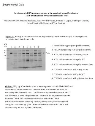
MW kDa 1 2 3 4 5 6 7 8 82 62 49 37 1. Purified His-tagged-polµ (positive control) 2. Poll overexpressing cells (negative control) 3. A’7H cells transfected with empty vector 4. A’7H cells transfected with polµ WT 5. A’7H cells transfected with polµ inactive form 6. C’10 cells transfected with empty vector 7. C’10 cells transfected with polµ WT 8. C’10Cells transfected with polµ inactive form Supplemental Data Involvement of DNA polymerase mu in the repair of a specific subset of DNA double strand breaks in mammalian cells Jean-Pascal Capp, François Boudsocq, Anne-Gaelle Besnard, Bernard S. Lopez, Christophe Cazaux, Jean-Sébastien Hoffmann and Yvan Canitrot. Figure S1: Testing of the specificity of the polµ antibody. Immunoblot analysis of the expression of polµ in stably transfected cells. Polµ Methods: Fifty µg of total cells extracts were separated on 10% SDS-PAGE and transferred to PVDF membrane. The membrane was blocked 1 h with 5% non fat dry milk diluted in TBS-T (0.5% tween 20) washed twice with TBS-T then incubated at room temperature for 1 hour with the polµ antibody (1/500) diluted in TBS-T. The membrane was washed twice with TBS-T and incubated with the secondary antibody (horseradish peroxidase (HRP)- conjugated anti-rabbit IgG) for 1 hour washed three times with TBS-T and revealed using the ECL system (Amersham).