Objective X-ray Image Display Evaluation (OXIDE)
Objective X-ray Image Display Evaluation (OXIDE). May 2014. Katrin Helbing / TSA / OSC / EBSP. Background.

Objective X-ray Image Display Evaluation (OXIDE)
E N D
Presentation Transcript
Objective X-ray Image Display Evaluation (OXIDE) May 2014 Katrin Helbing / TSA / OSC / EBSP
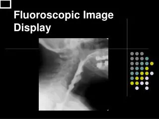
Background An inherent component of aviation security at our nation’s airports is the transportation security officers’ (TSOs) ability to detect potential threats in carryon and checked baggage. TSOs review displayed X-ray (2D) and CT (3D) images of passenger’s bags. Visual inspection of these images plays a large part in the security effectiveness.
The Issue… • There are currently no objective methods to quantify X-ray or CT image quality for both fixed and moving images as they apply to security screening. • Image quality is not tied to operator performance or capabilities, nor is it tied to detection of various threat components. • Image quality is currently assessed by running a ‘test kit’ (ASTM F792) through the x-ray machine, and ‘seeing’ what the smallest resolvable element is on the screen. ASTM F792 Step Wedge
The issue… (cont’d) • Problems with this method include: • Subjectivity; • As observer/assessor becomes more familiar with the test kit and resultant images, ‘performance’ improves (this likely stems from learning, optical illusions, and expectations as to what ‘should’ be seen); • Quality of the displayed image varies depending upon orientation of the test kit within the x-ray tunnel and image tools use. Therefore, assessed “image quality” is dependent on the presentation/position of the test article; • The test kit, although standardized across the industry, does not include ‘realistic’ test articles, and the resulting images are not representative of operations; • Image must be stationary for assessment. Can be used for fixed images only. TSA is implementing “continuous belt” operations - image will be moving as the TSO review it. There is a need to determine image quality of moving images as viewed by the operator.
A Solution… • DHS S&T Human Factors requested ideas for Small Business Innovation Research (SBIR) topics • Submitted need for Objective, Quantitative Image Quality Measurements and Metrics for Screener Imaging Technologies Objective, Quantifiable Result Tool, Measurement, Analysis Image # • Limitations: • Measure image quality from perspective of typical user (off of the display), not based solely on system-generated image data • No access or interface with vendor software or other system components • No access to vendor-proprietary algorithms • Initial CONOPs for laboratory use 20/20 9876 3.14 X magic happens…
SBIR Awarded • Phase 1: Develop a tool that provides an objective metric to quantify image quality as it is presented on the screen. Image quality should be measurable for both fixed and moving imagery. • Selected 3 vendors from 10 submittals (Sept. 2012) • 3 prototype systems were developed (April 2013) • Phase 2: Tie image quality metrics to human performance and capabilities as they relate to X-ray and CT image review (7/2013 – 7/2014) • Down selected to 1 vendor to complete project: Charles River Analytics, Inc. • Refine tool, automate process, develop GUI • Contract w/ Draper Labs HF experts to correlate metrics with human performance • Phase 3: Technology transition and commercialization (7//2014 – 7/2015) • Define additional display technologies • Refine tool use for continuous image quality monitoring / degradation
OXIDE Goals Objective X-ray Image Display Evaluation (OXIDE) • Key research goals include: • Development of novel edge-based image measures for effectively describing complex structures in x-ray imagery • Development of techniques for robustly modeling the relationship between performance, as it relates to the detection and identification of test kit items, with functional image quality ratings and human performance • Uses COTS hardware with custom algorithms • The achievement of these research goals will enable the eventual design, development, and deployment of a robust image quality measurement system
OXIDE Technical Approach • Use detectors trained on real collected images of a custom test kit to objectively inform a predictive algorithm • Algorithm assesses the presence and magnitude of different types of image degradations on novel imagery that may or may not contain the test kit • These degradations alter image quality, which determines if the image contains sufficient information for a detection to be made • Assess whether the image has sufficient quality to enable detection by a human operator • Approach will robustly handle changes to images caused by object orientation, glare, blur (including motion blur), projective distortion and noise • Can be applied to fixed and moving imagery
OXIDE Phase I Results • Semantic Interpretation • Performance Prediction • Evaluation • System Architecture • Data and Hardware • Test Kit Item Detection
Phase I: Image Degradation Metrics Motion Blur Noise Contrast
OXIDE Phase II • Requirements Elicitation • Data Collection • Hardware Prototype • Object Detection Refinement • Human Performance Model • Image Metrics Expansion • Software Interfaces • System Evaluation • Are there any restrictions on computing hardware or certifications required? • E.g., tablet, laptop • What are high-priority capability needs? • E.g., image quality factor prediction vs. general image scores • What set of image quality factors are most desirable beyond contrast, noise, and motion blur? • E.g., compression artifacts, aliasing, glare
Phase II: General Image Score (GIS) Lowest GIS • OXIDE measures the image, and calculates a General Image Score (GIS). • GIS is a single continuous value that objectively quantifies functional image quality • To begin validation of the GIS, degraded pristine imagery with varying levels of blurand other factors, sorted the images according to GIS. • GIS matches up with a human’s subjective ranking Highest GIS
Phase II: Auto-Calibration • Developed software tool for rapidly performing auto-calibration of the OXIDE camera system. • Calibration includes corrections for nonlinear deformations, such as rotation or perspective skew.
Phase II: GUI GIS • Graphical User Interface developed • Open File • Open Camera Stream • New General Image Score • Auto-Calibrate • View Image Statistics • Start/Stop Recording • Prototype hardware and software developed and delivered
Phase II: Next Steps A B • Correlate General Image Score (GIS) with human performance (pairwise comparisons) • TSIF Demo
What Next? • Interest in determining impact of X-ray and CT image quality on operator thereat detection performance. • Need to link image quality requirements to threat detection performance needs.