Download
1 / 25
250 likes | 467 Vues
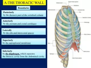
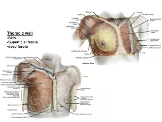
Muscles of Thoracic Wall. Muscles of the thorax consist of intercostals and Diaphragm. Intercostals are arranged as three layers between the ribs. Diaphragm closes the thoracic outlet and separates thoracic cavity from abdominal cavity. The Three layers of Intercostal Muscles are.

E N D
Muscles of the thorax consist of intercostals and Diaphragm. Intercostals are arranged as three layers between the ribs. Diaphragm closes the thoracic outlet and separates thoracic cavity from abdominal cavity.
The Three layers of Intercostal Muscles are External Layer: External Intercostal Internal Layer: Internal Intercostal Innermost Layer: AnteriorlyTransversus Thoracic Posteriorly Innermost intercostals and subcostal
External Intercostal Muscles There are 11 pairs of external intercostal muscles. They form the most superficial layer. Their fibres run infero-anteriorly(downwards and forward) from inferior border of rib above to the superior border of rib below. The muscle extends forwards to the costal cartilage where it is replaced by an aponeurosis, the anterior/external intercostal membrane.
Internal Intercostals • These muscles form the intermediate layer. Its fibers are directed downward and backward(infro-posteriorly) from the subcostal groove of the rib above to the upper border of the rib below. They are attached in form of aponeurosis called posterior intercostal membrane.
Innermost Intercostals These muscles form the deepest layer and correspond to transversus abdominis muscle in the anterior abdominal wall. They are related internally to endothroacic fascia and parietal pleura, externally to intercostal nerves and vessels.
Transversus Thoracis • These muscles originate from the lower part of the manubrium and xiphoid process, attaching to the costal cartilages of ribs 2-6. They are continuous with the transversus abdominus inferiorly.
Actions • External Intercostals increase the volume of the thoracic cage during forced inspiration. • Internal Intercostals decrease the volume of the thoracic cage during forced expiration. • Innermost Intercostals Same as Internal Intercostals
Neurovascular Bundle Contains arteries, veins and nerves.
Intercostal Arteries • Posterior Intercostal arteries of the first two spaces are branches of superior intercostal artery which is a branch of costocervical trunk of subclavian artery. Posterior Intercostal arteries of lower nine spaces are branches of thoracic aorta.
Anterior intercostal Arteries • Of first six spaces are branches of internal thoracic artery(arises from 1st part of subclavian). The anterior intercostal arteries of lower spaces are branches of musculophrenic artery, one of terminal branches of Internal Thoracic.
Veins • Anterior intercostal veins drain into internal thoracic and musculophrenic veins. • Posterior intercostal veins drain into azygous or hemi-azygous veins.
Nerves • Intercostal nerves are anterior rami of first 11 thoracic spinal nerves. • Each intercostal nerve enters the intercostal space between the parietal pleura and the posterior intercostal membrane. It then runs forward inferiorly to the intercostal vessels in the subcostal groove between the interna and innermost intercostal muscles. The first six I/C nerves are distributed within their I/C spaces.
Every I/C nerve is connected via a ganglion to the sympathetic trunk. • Collateral branches are given by all I/C nerves • Lateral Cutaneous Branch • Anterior Cutaneous Branch • Muscular Branches