Recombinant DNA Technology
340 likes | 456 Vues
Learn how to locate a specific gene in a vast genome through PCR, molecular cloning, and hybridization. Discover the benefits and limitations of Polymerase Chain Reaction (PCR) and its cycle steps. Explore applications, electrophoresis, and genetic markers like SSRs.

Recombinant DNA Technology
E N D
Presentation Transcript
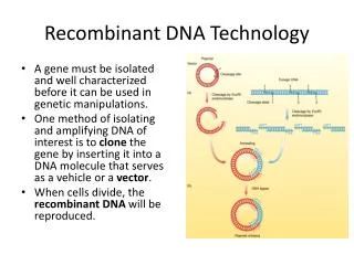
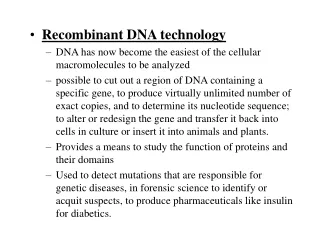
Needles in Haystacks • How to find one gene in large genome? A gene might be 1/1,000,000 of the genome. Three basic approaches: • 1. Polymerase chain reaction (PCR). Make many copies of a specific region of the DNA. • 2. cell-based molecular cloning: create and isolate a bacterial strain that replicates a copy of your gene. • 3. hybridization: make DNA single stranded, allow double strands to re-form using a labeled (e.g. radioactive) version of your gene to make it easy to detect.
Polymerase Chain Reaction • Based on DNA polymerase creating a second strand of DNA. • Needs template DNA and two primers that flank the region to be amplified. Primers are short (generally 18-30 bases) DNA oligonucleotides complementary to the ends of the region being amplified. • DNA polymerase adds new bases to the 3' ends of the primers to create the new second strand. • go from 1 DNA to 2, then 4, 8, etc: exponential growth of DNA from this region • A key element in PCR is a special form of DNA polymerase from Thermusaquaticus, a bacterium that lives in nearly boiling water in the Yellowstone National Park hot springs. This enzyme, Taq polymerase, can withstand the temperature cycle of PCR, which would kill DNA polymerase from E. coli. • advantages: • rapid, sensitive, lots of useful variations, robust (works even with partly degraded DNA) • disadvantages: • Only short regions (up to 2 kbp) can be amplified. • limited amount of product made
PCR Cycle • PCR is based on a cycle of 3 steps that occur at different temperatures. Each cycle doubles the number of DNA molecules: 25-35 cycles produces enough DNA to see on an electrophoresis gel. Each step takes about 1 minute to complete. • 1. Denaturation: make the DNA single stranded by heating to 94oC • 2. Annealing: hybridize the primers to the single strands. Temperature varies with primer, around 50oC • 3. Extension: build the second strands with DNA polymerase and dNTPs: 72oC.
DNA Amplification in PCR • original DNA: very long molecules with neither end well defined. Number stays constant in the PCR reaction: no new ones are made. • initial PCR product made from original DNA: has one end defined by the primer, but the other end is not well defined. Copy number grows linearly. • all other PCR products have 2 ends defined by the primers, so they have a constant length and can be easily detected by electrophoresis. Copy number grows exponentially.
Electrophoresis • Separation of charged molecules in an electric field. • Nucleic acids have 1 charged phosphate (- charge) per nucleotide. means constant chare to mass ratio. Separation based (mostly) on length: longer molecules move slower. • Done in a gel matrix to stabilize: agarose or acrylamide. • average run: 100 Volts across a 10 cm gel, run for 2 hours. • Stain with ethidium bromide: intercalates between DNA bases and fluoresces orange. • Run alongside standards of known sizes to get lengths
PCR Applications • RT-PCR: use reverse transcriptase to convert messenger RNA into DNA, then amplify it with PCR. • Anchor-primed PCR: use one sequence-specific primer and use a set of random primers for the other end. For example: 3’ RACE-PCR (Rapid Amplification of cDNA Ends) uses an oligo-dT primer to bind to the poly A tail of mRNA and a universal primer for the internal region. • Adding linkers to the primers puts them into the amplified DNA. Useful for cloning or further PCR.
Allele-Specific PCR • For base change mutations (single nucleotide polymorphisms). • Use a primer whose 3’ base matches the mutation. Will amplify one allele but not the other because the 3’ end is not paired with the template in the wrong allele.
SSR Genetic Markers • . Microsatellites (Simple Sequence Repeats: SSRs). Used for mapping the human genome--the main marker system used today. • SSRs are short (2-5 bases) sequences that are repeated several times in tandem: TGTGTGTGTGTG is 6 tandem repeats of TG. • SSRs are found in and near many genes throughout the genome--they are quite common and easy to find. • During normal replication of the DNA in the nucleus, DNA polymerase sometimes slips and creates extra copies or deletes a few copies of the repeat. • This happens rarely enough that most people inherit the same number of repeats that their parents had (i.e. SSRs are stable genetic markers), but often enough that numerous variant alleles exist in the population. • Mapping SSRs is a matter of having PCR primers that flank the repeat region, then examining the PCR products on an electrophoresis gel and counting the number of repeats. • SSRs are co-dominant markers: both alleles can be detected in a heterozygote. • If an SSR is a 3 base repeat within the coding region of a gene, it will create a tandem array of some amino acid. Certain genetic diseases, most notably Huntington's Disease, are caused by an increase in the number of repeats: once the number gets high enough the protein functions abnormally, causing neural degeneration. Such SSRs are called "tri-nucleotide repeats" or TNRs.
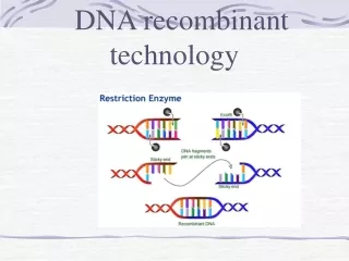
Cell-Based Molecular Cloning • The original recombinant DNA technique: 1974 by Cohen and Boyer. • Several key players: • 1. restriction enzymes. Cut DNA at specific sequences. e.g. EcoR1 cuts at GAATTC and BamH1 cuts at GGATCC. • Used by bacteria to destroy invading DNA: their own DNA has been modified (methylated) at the corresponding sequences by a methylase. • 2. Plasmids: independently replicating DNA circles (only circles replicate in bacteria). Foreign DNA can be inserted into a plasmid and replicated. • Plasmids for cloning carry drug resistance genes that are used for selection. • Spread antibiotic resistance genes between bacterial species • 3. DNA ligase. Attaches 2 pieces of DNA together. • 4. transformation: DNA manipulated in vitro can be put back into the living cells by a simple process . • The transformed DNA replicates and expresses its genes.
Plasmid Vectors • To replicate, a plasmid must be circular, and it must contain a replicon, a DNA sequence that DNA polymerase will bind to and initiate replication. Also called “ori” (origin of replication). • Replicons are usually species-specific. • Some replicons allow many copies of the plasmid in a cell, while others limit the copy number or one or two. • Plasmid cloning vectors must also carry a selectable marker: drug resistance. Transformation is inefficient, so bacteria that aren’t transformed must be killed. • Most cloning vectors have a multiple cloning site, a short region of DNA containing many restriction sites close together (also called a polylinker). This allows many different restriction enzymes to be used. • Most cloning vectors use a system for detecting the presence of a recombinant insert, usually the blue/white beta-galactosidase system.
Basic Cloning Process • Plasmid is cut open with a restriction enzyme that leaves an overhang: a sticky end • Foreign DNA is cut with the same enzyme. • The two DNAs are mixed. The sticky ends anneal together, and DNA ligase joins them into one recombinant molecule. • The recombinant plasmids are transformed into E. coli using heat plus calcium chloride. • Cells carrying the plasmid are selected by adding an antibiotic: the plasmid carries a gene for antibiotic resistance.
DNA Ligase in Action! I hope
Cloning Vector Types • For different sizes of DNA: • plasmids: up to 5 kb • phage lambda (λ) vectors: up to 50 kb • BAC (bacterial artificial chromosome): 300 kb • YAC (yeast artificial chromosome): 2000 kb • Expression vectors: make RNA and protein from the inserted DNA • shuttle vectors: can grow in two different species
Lambda-based vectors • Phage lambda can do 2 different things when it enters the cell: • lytic cycle: it can start reproducing itself immediately, which produces about 200 new phage in 15 minutes and kills the cell • lysogenic cycle: the lambda DNA can integrate into the host chromosome and remain dormant for many generations. When given the proper signal, the integrated DNA (prophage) leaves the chromosome and enters the lytic cycle. • Lambda is about 50 kb long, and the central 20 kb is only used for lysogeny; it can be replaced by foreign DNA. • Ligation of arms with insert using DNA ligase • Packaged into phage particles in vitro using extracts from cells that have contain pieces of the phage heads. • Use these phage to infect new E. coli. • Cosmids are similar to phage vectors: use lambda, but remove all but the ends (cos sites), ori, and selectable marker. Package in vitro--becomes a large (50 kb) plasmid in the E. coli.
Bacterial Artificial Chromosomes • Based on the F plasmid that confers the ability to conjugate. • Low copy number plasmids (usually 1 per cell), which prevents crossing over between repeated sequences in the insert DNA • But, low copy number also means low DNA yield. • Transformed into E. coli using electroporation, subjecting the bacteria to a high voltage electrical field. • BACs are currently the most common vector for large inserts such as eukaryotic genome projects.
Yeast Artificial Chromosomes • A linear chromosome, has centromere, telomeres, ARS (autonomously replicating sequence), selectable marker for yeast (uracil or tryptophan biosynthesis genes usually). • Also has E. coli ori and selectable marker: you can grow the vector itself in E. coli • Then purify it, ligate in foreign DNA, transform into yeast.
Expression Vectors • Various types: • RNA only: use a vector that has a phage T7 promoter in front of the cloning site, and an inducible T& polymerase gene. Induction by the lac operon repressor gene and the synthetic inducer IPTG (isopropyl thiogalactoside). • polypeptide or fragments of polypeptides: can be produced in E. coli using a ribosome binding site in addition to the promoter. Need to use the correct reading frame. • can also be done as a fusion protein (your protein fused to a marker protein) for easy detection or purification • post-translationally modified or intron-spliced protein: needs to be expressed in eukaryotic cells. Needs eukaryotic promoter and polyadenylation (poly-A addition) signals, plus a selectable marker that works in eukaryotes (since most antibiotics are specific for prokaryotes).
Example Expression Vector • For eukaryotic expression, this vector (from Invitrogen) has a cauliflower mosaic virus promoter (PCMV), a bovine growth hormone polyadenlyation site (BGHpA). • The DNA inserted at “hORF” gets fused to a short peptide called an epitope, for which very specific anitbodies exist. It also gets fused to 6 histidines, which allow easy purification on a column that has nickel ions bound to it (an affinity tag). • For growth in mammalian cells, it has an SV40 viral origin of replication (SV40ori), and a zeocin resistance gene (Zeocin, with SV40 promoter/enhancer and SV40 poly A site). • For growth in E. coli it has the ColE1 replicon. Zeocin works as a selectable marker in baceria as well as in eukaryotic cells. • There is also a T7 promoter for making RNA from the inserted gene, and an f1 origin of replication for making single stranded DNA (useful for sequencing).
Sources of DNA to Clone • Genomic DNA: cut up whole genome and clone small pieces. Advantage is, you get everything. Disadvantage is, a lot of it is junk. • Two general methods: • 1. randomly shear DNA into small pieces, then ligate linkers to the ends: oligonucleotides that contain a useful restriction site. • 2. partially digest the DNA with a restriction enzyme that has a 4 base recognition site. These sites will appear at random every 256 (44) base pairs. Take long pieces. • cDNA: DNA copy of mRNA, made with reverse transcriptase. Advantage: you just get the expressed genes. Disadvantages: you don't get control sequences or introns, and frequency depends on level of expression. • Synthetic DNA: synthesized de novo (for example multiple cloning sites or linkers), or made by PCR
cDNA Synthesis • use oligo-dT primer, which binds to poly-A tail. • make the first DNA strand from the RNA using reverse transcriptase
More cDNA Synthesis • Remove the RNA with heat or alkali. • The 3’ end spontaneously forms a small hairpin. • Extend the hairpin with DNA polymerase • Cut eh loop with S1 nuclease (which cuts at unpaired bases) • Attach synthetic linkers with DNA ligase and clone into a vector.
Libraries • A large number of clones, often pooled together (so you have to fish out the one you want), but sometimes ordered. • Genomic library vs. cDNA. • Genomic uses enough input DNA to cover the genome 5-10x, so chance fluctuations don't prevent all sequences from being cloned. Repeat sequence DNA is a problem. • cDNA libraries are usually made from single tissues: expression varies between tissues. Large difference in expression levels, often compensated for by normalizing the library: trying to equalize copy number of different sequences. • detection of clones containing specific genes is generally by hybridization with labeled probes. It can also be done using antibodies if the genes in the library are being expressed.
Hybridization • The idea is that if DNA is made single stranded (melted), it will pair up with another DNA (or RNA) with the complementary sequence. If one of the DNA molecules is labeled, you can detect the hybridization. • Basic applications: • Southern blot: DNA digested by a restriction enzyme then separated on an electrophoresis gel • Northern blot: uses RNA on the gel instead of DNA • in situ hybridization: probing a tissue • colony hybridization: detection of clones • microarrays
Labeling • Several methods. One is random primers labeling: • use 32P-labeled dNTPs • short random oligonucleotides as primers (made synthetically) • single stranded DNA template (made by melting double stranded DNA by boiling it) • DNA polymerase copies the DNA template, making a new strand that incorporates the label. • Can also label RNA (sometimes called riboprobes), use non-radioactive labels (often a small molecule that labeled antibodies bind to, or a fluorescent tag), use other labeling methods.
Hybridization Process • All the DNA must be single stranded (melt at high temp or with NaOH). Occurs in a high salt solution at say 60oC. Complementary DNAs find each other and stick. Need to wash off non-specific binding. • Stringency: how perfectly do the DNA strands have to match in order to stick together? Less than perfect matches will occur at lower stringency (e.g. between species). Increase stringency by increasing temp and decreasing salt concentration. • Rate of hybridization depends on DNA concentration and time (Cot), as well as GC content and DNA strand length. • Autoradiography. Put the labeled DNA next to X-ray film; the radiation fogs the film.
Southern Blot • Used to detect a specific DNA sequence in a complex mixture, such as genomic DNA • Cut DNA with restriction enzyme, then run on an electrophoresis gel. • Suck buffer through the gel into a nitrocellulose membrane. The buffer goes through but the DNA sticks to the membrane. • Fix the DNA to the membrane permanently with UV or heat • Hybridize membrane to a radioactive probe, then detect specific bands with autoradiography. • Northern blot uses RNA instead. RNA must be denatured so the distance it migrates on the gel is proportional to its length: put formaldehyde in the gel.
Restriction Fragment Length Polymorphisms • RFLPs: the first DNA-based genetic mapping technique. Advantage: every individual has many variations in their DNA, so you don’t need a special set of marker mutations. Also, the markers are co-dominant so you can accurately determine the genotype. • Probe is a fragment of a cloned gene (labeled). • Genomic DNA is cut with a restriction enzyme. • Polymorphic sites: the restriction site is present in some individuals but not in others (due to mutation). But, even if one site is missing, there will be another restriction site a little further away (a restriction enzyme with a 6 base site cuts on the average every 46 = 4096 bp). • Then do a Southern blot and autoradiography.
Colony Hybridization • Bacterial colonies (or phage plaques) containing recombinant DNA are grown on agar, then blotted to nitrocellulose and hybridized as with Southern blots. • The colonies on the agar plates stay alive, and once the correct colony has been detected, it can be picked and grown up for further work.
In Situ Hybridization • Using tissues or tissue sections. • Often done with non-radioactive probes because the high energy of 32P emission gives an imprecise view of where the hybridization is. • Counterstain the tissue so non-hybridizing parts are visible.
Microarrays • Place probes from many different genes on a glass microscope slide, then hybridize to cDNA made from messenger RNA isolated from a tissue. You see which genes are active in that tissue. • Mostly done with 60mers: 60 bases long, synthetic oligonucleotides, made using sequence information from the genes. • cDNA is fluorescently labeled • Often 2 conditions are compared (control and experimental), using red and green fluorescent tags. • Semi-quantitative • Can also be used to screen for DNA mutations.