Download

1 / 8
80 likes | 377 Vues
Ghimenton C 1 , Eccher A 1 , Brunelli M 2 , Gobbo S 2 , Parisi A 1 , Martignoni G 2 , Turazzi S 3 , Chilosi M 2 , Menestrina F 2 . 1 Anatomia Patologica, Ospedale Civile Maggiore, Verona 2 Dipartimento di Patologia, Università di Verona 3 Neurochirurgia, Ospedale Civile Maggiore, Verona.

E N D
Ghimenton C1, Eccher A1, Brunelli M2, Gobbo S2, Parisi A1, Martignoni G2, Turazzi S3, Chilosi M2, Menestrina F2. 1 Anatomia Patologica, Ospedale Civile Maggiore, Verona 2 Dipartimento di Patologia, Università di Verona 3 Neurochirurgia, Ospedale Civile Maggiore, Verona Extramedullary hematopoiesis as a potential pitfall for meningeal mass in patients with myelofibrosis
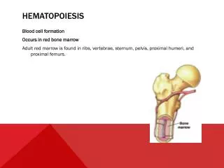
Introduction • Myelofibrosis leads toineffective erythropoiesis • Extramedullary hematopoiesis characteristically • develops predominantly in the spleen and liver • The central nervous system is rarely affected and the • spinal canal and the meninges are preferred locations
Case report • 63 yrs woman with history of worsening headaches and • documented bone marrow fibrosis. • MR imaging revealed a bilateral, frontal-solid-lesion, • attached to the dura, that was hyperintense to contrast. • The patient underwent a frontal craniotomy • for suspected meningioma and the lesion was removed • with its dural implant. • Grossly the tumor resection measured in • aggregate cm 4,5x3,5x3.
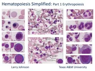
Histological examination revealed multiple foci of hematopoietic cells of erythroid, myeloid and megakaryocytic lineages confirmed with stains for glycophorin, myeloperoxidase and Factor VIII.
The hematopoietic cells were of course immunonegative for pankeratin and glial fibrillary acidic protein. The diagnosis was meningeal EH. AE1/AE3 GFAP
Discussion • The presence of EH loci in primary brain tumors • is fairly rare • Extramedullary myeloid metaplasia should be ruled out • in patients experiencing idiopathic myelofibrosis and • considered as a differential diagnosis in brain lesions
References • A meningioma with islets of extramedullary myeloid metaplasia: • case report. • Zona G. et al, Neurosurgery 2007. 2. Intracranial extramedullary hematopoiesis associated with pilocytic astrocytoma: a case report Beckner E. et al, Acta Neuropathology 2003.