Download
1 / 60
610 likes | 659 Vues
Explore the male and female reproductive systems, from anatomy to hormonal regulation, to understand fertility and reproductive health. Learn about spermatogenesis, ova production, and factors influencing reproductive functions.

E N D
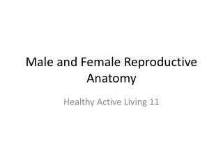
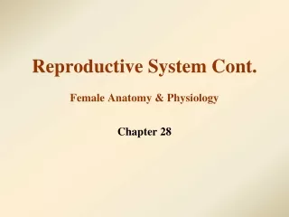
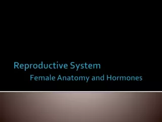
Anatomy and Physiology of the male and female Reproductive System
Broad Objective • To acquire knowledge on anatomy and physiology of reproductive system and develop skills and attitudes in order to manage reproductive health problems efficiently.
Specific objectives • Describe anatomy and physiology of the male and female reproductive system • Describe the process of fertilization and implantation • Describe the functional anatomy of the breast • Discuss pubertal/secondary characteristics changes in both male and female adolescents • Explain developmental anomalies in both male and females
Anatomy and physiology of male and female reproductive system • The normal physiological functioning of the Reproductive System depends on stimulation from the brain. The stimulation can be in the form of hormones or impulses. • The major parts of the brain involved in the reproductive system function include:- Cortical areas, Cingulate ganglion, Caundate nuclei, Frontal cortex, Inferior temporal cortex • The major glands involved in hormonal production are Hypothalamus and pituitary and the gonads
Contd… • The hormones involved in reproductive system regulation include gonadotrophins (Follicle Stimulating Hormone and Leutinizing Hormone), which are produced by the pituitary gland. • The production of the gonadotriphin hormones is regulated by gonadotrophin Releasing hormone (GnRH) that produced in the hypothalamus. • The gonadotrophins regulate the production of gonadal hormones.
Contd… • The gonadal hormones include oestrogen and progesterone produced by the ovaries and testosterone produced by the testis. • The gonadotrophins also regulate the spermatogenesis and development of the ovum. • Hormonal production is regulated by a negative feedback mechanism.
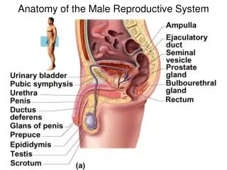
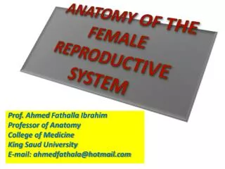
Anatomy and physiology of male reproductive system Lateral view of the male reproductive system
Functions of the male reproductive system structures • Penile shaft – is the organ for sexual intercourse and for passing urine. • Testis – the male sexual gland. It produces sperms and testosterone hormone. • Prostate and seminal vesicle – produces seminal fluid, which is necessary for sperm mobility. • Urethral duct – the canal that ensures passage of sperms and urine.
Contd… • Vas deferens- Referred to as the spermatic cord or epididymis. It is a channel for sperms from the testis to the urethra. Sperm maturation takes place in this channel. This is the duct that is severed during vasectomy. • Cowper’s gland Referred to as bulbo-urethral gland. It produces a fluid, which protects the sperm during transportation by neutralizing the acidity in the urethra.
Contd… • Scrotum This is a sheath surrounding the testis. Apart from protecting the testis, it also regulates testicular temperature as well as acting as an erogenous zone.
Physiology of male reproductive system • Control of male gonads is by hormones as outlined above. • FSH works on the seminiferous tubes to bring about spermatogenesis while • LH acts on interstitial cells to produce testosterone. • Testosterone is responsible for the secondary sexual characteristics.
Spermatogenesis • Spermatogenesis begins at puberty and continues throughout adult life. • Whereas only few hundred ova are liberated for fertilization during the life span of a woman, billions of spermatozoa are formed in the testis starting from puberty and continuing until old age. • Although it takes only one spermatozoon to fertilize the ovum, there are normally about 20-100 million sperms per millilitre of seminal fluid. • The large number of spermatozoa is necessary in order to overcome the difficulties, which they encounter during the journey to the ovum in the uterine tube.
Contd… • Only a few thousand are able to reach the site of fertilization. The majority are destroyed by the often-unfavourable chemical medium found in the vagina, by phagocytosis and by absorption by the uterine wall. • The Process of spermatogenesis takes about 70 days and is continuous (after vasectomy one has to use a condom for about 90 days since sperms are still present in the vas deferens for a full sperm cycle)
Factors that can affect spermatogenesis • Undescended testis (cryptochirdism) • Increased scrotal temperature (can be caused by wearing tight underwear, scrotal varicose veins) • Infection of the scrotum and testis e.g. childhood mumps, epididymorchitis • Tortion of testis • Drugs and alcohol
Anatomy and physiology of female reproductive system Diagram of the female external genitalia
Functional anatomy of the female reproductive organs • Mons pubis – is the pad of fat, which is covered by hair at the onset of puberty. Normal pubic hair in the female is distributed in an inverted triangle at the mons pubis. • Labia majora – these are two large folds of skin which cover the external genitalia. • Labia minora – these are two smaller folds of skin located beneath the labia majora. As they join in the upper part they form the hood of the clitoris. During sexual arousal, they secrete a thick white substance, which assist in sexual intercourse.
Contd… • Clitoris – it is an erectile tissue that corresponds to the male penis. Its only known role is erotic. It is always excised during female circumcision (Clitoreidectomy) Vestibule – is the area between the two labia minorawhere the vagina, urethral orifice, and the skenes glands are located. • Vagina – the vagina is the canal that connects the uterus to the external genitalia. It is main functions are to allow sexual activity and vaginal birth. Its walls are made of muscle and mucosal layer. The muscles are arranged in a circular manner, which allows the vaginal walls to expand and contract. The rugae in the mucosal layer are important for sensation during sexual intercourse.
Contd… • Uterus - This includes the uterine body and the cervix. The function of the uterus is to accommodate the pregnancy. The activity of the ovarian hormones on the uterus leads to the formation of menstrual blood. • Cervix –This is the lower part of the uterus that opens into the vagina. Capacitiation of the sperm takes place in the cervix. This enables the sperm to travel up the uterus and to fertilize the ova. The cervix also produces mucus, which closes the cervix and prevents germs from entering the uterus. The cervical mucus plug thickens when one is using contraceptives thus enhancing efficacy. At the onset of labour, the cervical mucus plug mixed with blood comes out as show.
Contd… • Cancer of the cervix is the commonest reproductive tract cancer in females in Kenya. • Ovaries – these are the female gonads that produce the ova and the female gonadal hormones (oestrogen and progesterone). • Fallopian tubes; – • They are the ducts in charge of collecting carrying and feeding the ovum until its arrival in the uterus. Usually fertilization takes place here. • They are easily damaged during pelvic infection leading to infertility. • Ectopic pregnancy in the tubes may occur as result of tubal infection and can be life threatening. • They are severed during bilateral tubal ligation.
Physiology of the female reproductive system • Effect / roles of hormones • Ovarian Cycle • Menstrual cycle • Fertilization and implantation
Effect of hormones on the ovaries • Control of female gonads is hormonal and cyclical. Maturation of the hormonal and gametogenic functions of the female gonads is initiated during puberty. • The cyclic nature of the menstrual cycle is determined at this time
Effect of hormones on the ovaries… • FSH causes maturation of graafian follicles and also stimulates the theca cells and granulosa cells to secrete oestrogen • LH causes release of the mature ovum leaving the corpus luteum • When ovulation does not occur, no corpus luteum is formed
Effect of hormones on the ovaries… • After ovulation, progesterone and minimal amounts of oestrogen are produced by the corpus luteum • During menopause there is atrophy of the ovaries with resultant cessation of ovulation and hormonal synthesis
Role of progesterone hormone • High levels of progesterone are required for the maintenance of a pregnancy to term • It stimulates the development of the breast alveoli and lobules, and supports lactation • Progesterone increases body temperature. This is the basis for using basal body temperature (BBT) in natural family planning • A high level of progesterone prevents ovulation • Some progesterone preparations are used to induce labour e.g misoprostol (cytotech)
Role of oestrogen • Responsible for the development of secondary sexual characteristics • Leads to the growth of the internal and external genital organs particularly the uterus, the vagina and the labia majora • Responsible for the feminine look and behaviour (feminising hormone) • Causes breast enlargement
Role of oestrogen… • It increases libido • It has a role in the cyclic changes in the endometrium that leads to menstruation • Large doses of oestrogen can lead to blood clotting problems (thromboembolic disorders, endometrial cancer, contraception among others • Oestrogen and progesterone work together to maintain the hormonal equilibrium
Ovarian Cycle • The ovarian cycle has two phases; 1) Follicular and 2) Luteal • The follicular phase begins from the first day of menstruation to the time of ovulation while the Luteal phase begins from ovulation until the start of the next menstruation.
Follicular phase • From the time of birth, there are many primordial follicles under the ovarian capsule. Each contains an immature ovum • At the start of each cycle, several of these follicles enlarge and a cavity forms around the ovum (antrum formation). • In humans, one of the follicles, (dominant follicle), in one ovary starts to grow rapidly on about the sixth day of the menstrual cycle, while the others regress (atretic follicles).
Follicular phase • It is not known how one follicle is singled out for development during this follicular phase of the menstrual cycle • When women are given highly purified human pituitary gonadotrophin preparations by injection, many follicles develop simultaneously
Ovulation • At the 14th – 16th day of the cycle, the dominant follicle ruptures, and the ovum is extruded into the abdominal cavity. This is the process of ovulation • The ovum is picked up by the fimbrial ends of the fallopian tubes (oviducts) • Fertilization normally occurs in the proximal end of the fallopian tubes. If fertilization does not occur, the ovum degenerates
Luteal phase • The follicle that ruptures at the time of ovulation develops to form the corpus luteum • Minor bleeding from the follicle into the abdominal cavity may cause peritoneal irritation and fleeting lower abdominal pain (“mittelschmerz”).
Luteal phase • If conception occurs the corpus luteum persist to produce progesterone, which maintains the pregnancy until the placenta takes over the progesterone production. • If there is no pregnancy, the corpus luteum begins to degenerate about 4 days before the next menses (day 24 of the cycle) and is eventually replaced by fibrous tissue, forming a corpus albicans.
Menstrual Cycle / Uterine Cycle • In many cultures a girl is considered as a woman at menarche. It has been noted that the age at menarche is decreasing but it is normally between 10-13 years • In warmer climates menarche starts at an earlier age • Variations in oestrogen and progesterons levels are responsible for the dramatic changes in the endometrium throughout the menstrual cycle
Menstrual Cycle / Uterine Cycle… • At the completion of the menstrual period the endometrium is only 1-2mm millimetres thick • Under the influence of increasing levels of oestrogen the endometrium increases in thickness until by day 12 of the cycle when its 10 to 12 mm thick • This growth results from an increase in epithelial and stroma cells of the superficial layer of endometrium
Menstrual Cycle / Uterine Cycle… • This is called the Proliferative Phase and is characterized by an increase in oestrogen receptor content and increases in size of the endometrial glands • As ovulation approaches, the progesterone receptor content of the endometrium increases • Within two days of ovulation the effect of rising levels of progesterone becomes apparent as the endometrium enters the Secretory Phase of the cycle
Menstrual Cycle / Uterine Cycle… • During this phase the mitotic activity in the epithelium ceases. The glands and blood vessels become dilated and tortuous • Glycogen accumulation in the endometrium reaches its peak level under the combined influence of oestrogen and progesterone. This process prepares the endometrium for embedding of the embryo • If fertilization does not occur the progesterone and oestrogen levels decline resulting in menstruation
Menstrual Phase • It is characterized by vaginal bleeding and lasts 2-7 days. It’s the terminal phase of the cycle and is the period during which the endometrium is shed down upto the basal layer together with blood from the capillaries and the unfertilised ovum
PROCESS OF FERTILIZATION AND IMPLANTATION • Fertilization • Following ovulation, the egg should be fertilized within approximately 12-24 hours • Fertilization occurs in the ampulla of the fallopian tube, after which a zygote is formed. The zygote then moves to the endometrium for implantation
PROCESS OF FERTILIZATION AND IMPLANTATION… • Implantation • Once the Zygote reaches the blastocyst stage (approximately five to six days after fertilization), it begins the process of implantation • Implantation takes place when the Zygote attaches or implants at the fundus of the uterus
THE BREAST • Anatomy of the breast • Although the breast is not located in the pelvic area, it forms part of the female reproductive system • During puberty in response to hormonal stimulation, the female breast begins to enlarge and mature
Anatomy of the breast… • The nipple • The central area through which the milk ducts open. • The areola • The circular dark area around the nipple. The skin of the areola contains small elevated nodules or bumps (Montgomery’s tubercles), beneath which lie the glands of Montgomery • These glands are responsible for lubrication of the nipple and help prevent nipple and areola cracks and fissures
Anatomy of the breast… • Mammary glands -Mammary glands are compound tubulo-alveolar glands. They have 15-20 lobes that radiate out from the nipple separated by collagenous connective tissue. -The lobes drain into lactiferous ducts, which expand to form a lactiferous sinus for milk storage.
Anatomy of the breast… • Fat -80-85 percent of normal breast is fat. In late pregnancy the fat is replaced by glandular or milk cells in readiness for lactation. The fat gives the breast its shape.
Anatomy of the breast… • Ligaments of Cooper -These are bands of fascia that run from the breast into the sub-cutenious tissues. They support the breast in its upright position. As woman grows older, the ligaments weaken leading to sagging of the breast. -These ligaments may be distorted by breast tumours resulting in skin dimpling (lemon peal appearance).
Stages of breast development • Stage I • Pre-pubertal state, elevation of papilla only • Stage II • Breast budding – palpable breast tissue, areola and nipple begin to enlarge • Stage III • Further enlargement of entire breast tissue
Stages of breast development… • Stage IV • Secondary mound of areola and papilla projecting above the breast tissue • Stage V • Secondary mound as a rose in stage IV disappears. Adult breast with typical contour
Functions of the breast • Aesthetic – for cosmetic purposes • Erogenic – for sensuality • Lactation – for milk production and breast feeding