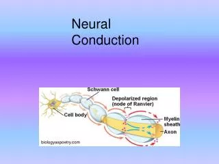
Neural Conduction
Neural Conduction. biologyaspoetry.com. Resting Membrane Potential. A cell’s membrane potential is the difference in the electrical potential ( ≈ charge) between the inside and outside of the cell. The inside of the neuron is negative with respect to the outside. -70mV.

Neural Conduction
E N D
Presentation Transcript
Neural Conduction biologyaspoetry.com
Resting Membrane Potential A cell’s membrane potential is the difference in the electrical potential (≈ charge) between the inside and outside of the cell. The inside of the neuron is negative with respect to the outside. -70mV Resting membrane potential is about –70mV. In its resting state, a neuron is said to be polarized.
Ionic Basis of the Resting Potential more sodium (Na+) and chloride (Cl-) outside than inside outside Na+ Cl- Na+ X- X- Na+ X- Na+ anions maintain overall charge neutrality on both sides of the membrane K+ Cl- K+ Na+ Cl- X- Na+ X- Cl- Na+ Na+ X- K+ A- cell membrane K+ Na+ A- A- K+ A- K+ Cl- K+ A- A- K+ K+ K+ A- A- Na+ A- more potassium (K+) inside than outside inside
Distribution of Ions 1 concentration gradient electrostatic pressure Factors contributing to even distribution of ions • Random motion: particles tend to move down their concentration gradient • Electrostatic pressure: like charges repel like; opposites attract Equilibrium potential The membrane potential at which there is no net flow of an ion (concentration and electrical gradients are equal and opposite)
Distribution of Ions 2 Factors contributing to uneven distribution of ions • Selective membrane permeability to certain ions • Sodium–potassium pumps Na+ Na+ K+ K+ biomhs.com www.eplantscience.com
K+ leaves the cell due to the concentration gradient K+channels + - K+ K+ K+ K+ enters the cell due to the electrical gradient Membrane Permeable Only to Potassium outside K+ K+ K+ K+ K+ Equilibrium Potential: -100mV K+ K+ K+ K+ K+ inside
Na+ leaves the cell due to the electrical gradient Na+channels - + Na+ enters the cell due to the concentration gradient Membrane Permeable Only to Sodium outside Na+ Na+ Na+ Na+ Na+ Na+ Na+ Na+ Na+ Na+ Na+ Na+ Na+ Equilibrium Potential: +40mV Na+ inside
Cl- leaves the cell due to the electrical gradient Cl- channels + - Cl- enters the cell due to the concentration gradient Membrane Permeable Only to Chloride outside Cl- Cl- Cl- Cl- Cl- Cl- Cl- Cl- Cl- Cl- Cl- Cl- Cl- Equilibrium Potential: -70mV Cl- inside
The Neuron at Rest: Tug-of-War At rest, the cell membrane is permeable to K+, Na+ and Cl- but is most permeable to K+ Thus, the resting membrane potential is drawn towards the K+ equilibrium potential and ends up at -70mV At rest, K+ leaks out, Na+ leaks in and Cl- is at equilibrium. Na+/K+ pumps transport 2 K+ in for every 3 Na+ they transport out to maintain the respective concentration gradients. academic.uprm.edu
Postsynaptic Potentials (PSPs) 1 Neurotransmitters bind at post-synaptic receptors. Chemical messengers make the postsynaptic membrane more or less permeable to specific ions. • Depolarizations (excitatory postsynaptic potentials, EPSPs) make the membrane potential more positive • Hyperpolarizations (inhibitory postsynaptic potentials, IPSPs) make the membrane potential more negative
Postsynaptic Potentials (PSPs) 2 PSPs are graded, rapid and decremental • Graded: amplitude proportional to intensity of stimulus (stronger stimuli = bigger PSPs) • Rapid: EPSPs and IPSPs travel quickly from their site of generation (receptor) to the soma • Decremental: they get smaller as they travel towards the soma www.studyblue.com receptor membrane potential millivolts soma time (ms)
PSPs and Neural Threshold In order to generate an action potential (AP; or to make a neuron “fire”), the threshold of activation (about -60 mV) must be reached near the axon hillock. One EPSP typically will not suffice — summation is needed. AP -60 threshold rest cnx.org
Types of Summation of PSPs Temporal summation Spatial summation integration of events happening at different places integration of events happening at different times
The Action Potential action potential Action potential (AP): a short-lasting event (~1 millisecond in duration) in which the membrane potential of a cell rapidly rises (from about -70 mV to around +20 mV) and falls. All-or-None: APs are not graded responses; their size (and velocity) is not related in any way to the intensity of the stimuli that elicit them. They occur to their full extent or do not occur at all (larger stimuli may elicit more APs than smaller stimuli).
Molecular Basis of the Action Potential APs are generated by special types of voltage-gated ion channels in the membrane of the axon (beginning at hillock). When threshold is reached, voltage-gated sodium channels open allowing an influx of sodium; as the AP nears its peak, these channels close and voltage-gated potassium channelsopen allowing potassium to flow out. These channels close more slowly resulting in a return overshoot. action potential
Dynamics of Voltage-Gated Channels The time-course of an AP can be divided into three phases: rising, falling and undershoot. The duration of these phases is dependent upon the opening and closing of the voltage-gated sodium and potassium channels.
Refractory Periods Refractory Period: Time during which the cell resists generating new APs. There are two periods: action potential Absolute (1 ms): impossible to initiate an AP because voltage-gated Na+ channels closed Absolute RF Relative RF Relative (2-4ms): an AP is possible for a stronger than normal stimulus (to overcome fact that voltage-gated K+ channels still open) Refractory periods prevent the backwards movement of APs and limit the rate of firing.
Conduction of Action Potentials The conduction of APs along an axon differs from that of PSPs in three ways: • Not graded: APs are all-or-none • Slow: channels open and close • Nondecremental: always same size Orthodromic conduction: in the natural direction, from cell body to terminal buttons Antidromic conduction: towards the cell body
Conduction of APs voltage-dependent Na+ channel (closed weakly) voltage-dependent K+ channel (closed) -70 -70 -70 -70 -70 -70 -70 -70 -70 -70 -70 -70 Axon Soma
1. depolarization from dendrites Conduction of APs -20 -25 -30 -35 -40 -60 -70 -70 -70 -70 -70 -70 Axon Soma 2. threshold is crossed
1. depolarization from dendrites Conduction of APs 3. sodium channels open 4. sodium flows in Na+ -20 -25 -30 -35 -40 -60 -70 -70 -70 -70 -70 -70 Axon Soma 2. threshold is crossed
Conduction of APs 4. sodium flows in Na+ Na+ -20 -25 -10 +5 +20 +20 -10 -30 -50 -70 -70 -70 Axon Soma 5. massive local depolarization 6. rapid passive propagation of depolarization (bidirectional)
Conduction of APs 7. sodium channels close strongly 8. potassium channels open and potassium flows out K+ Na+ -20 -25 -10 +5 +20 +20 -10 -70 -70 -70 -30 -50 Axon Soma 5. massive local depolarization
Conduction of APs 8. potassium channels open and potassium flows out 10. potassium channels close K+ Na+ -20 -25 -30 -30 -70 -80 -70 -70 -70 -70 -50 -40 Axon Soma 9. massive local repolarization
11. threshold crossed Conduction of APs K+ Na+ -20 -25 -30 -30 -70 -80 -70 -70 -70 -70 -50 -40 Axon Soma 6. rapid passive propagation of depolarization (bidirectional)
Action Potential Propagation APs do not travel continuously down the axon, but are triggered anew at each portion of the membrane that has voltage-gated channels. Saltatory conduction: in myelinated axons, APs “hop” from one node of Ranvier to the next. Speeds velocity from about 1-10 meters/second to up to 150 m/s.
Action Potential Blockers • The propagation of APs can be blocked or reduced by toxins or demyelinating diseases. • Sample toxins: • Na+ channel: tetrodotoxin from puffer fish • K+ channel: noxiustoxin from scorpions • Demyelination diseases: • CNS: Multiple Sclerosis • PNS: Guillian-Barre syndrome