Hepatocyte Proliferation Analysis in Non-Parabiotic and Parabiotic Mice Using BrdU Staining
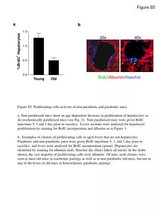
This figure illustrates the differences in hepatocyte proliferation between non-parabiotic and parabiotic mice of varying ages. Non-parabiotic mice displayed an age-dependent decline in hepatocyte proliferation, similar to their isochronic parabiotic counterparts. BrdU injections were administered prior to sacrifice, allowing for the analysis of proliferating cells in liver sections. Clusters of non-hepatocyte proliferating cells were identified, particularly in older mice. Significant observations include the prevalence of BrdU incorporation in albumin-positive cells and the absence of this condition in heterochronic parabiotic pairings.

Hepatocyte Proliferation Analysis in Non-Parabiotic and Parabiotic Mice Using BrdU Staining
E N D
Presentation Transcript
Figure S5 BrdU/Albumin/Hoechst a b 20x 40x Figure S5 Proliferating cells in livers of non-parabiotic and parabiotic mice. a, Non-parabiosed mice show an age-dependent decrease in proliferation of hepatocytes as do isochronically parabiosed mice (see Fig. 3). Non-parabiosed mice were given BrdU injections 5, 3 and 1 day prior to sacrifice. Livers sections were analyzed for hepatocyte proliferation by staining for BrdU incorporation and albumin as in Figure 3. b, Examples of clusters of proliferating cells in aged livers that are not hepatocytes. Parabiotic and non-parabiotic pairs were given BrdU injections 5, 3, and 1 day prior to sacrifice, and livers were analyzed for BrdU incorporation (green). Hepatocytes are identified by staining for albumin (red). Hoechst dye (blue) labels all nuclei. In the fields shown, the vast majority of proliferating cells were albumin-. Of note, such clusters were seen in most old mice in isochronic pairings as well as in non-parabiotic old mice, but not in any of the livers of old mice in heterochronic parabiotic pairings.