Download
1 / 40
400 likes | 565 Vues
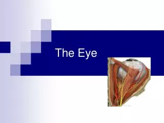
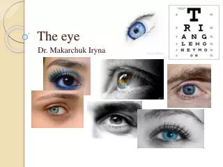
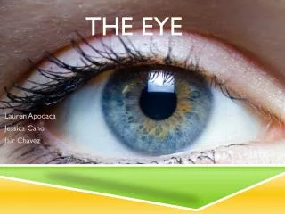
The Eye. Lauren Apodaca Jessica Cano Jair Chavez. Structure of the Eye. Outer Tunic Middle Tunic Inner Tunic. Outer Tunic. Made up of: - Cornea : clear avascular surface that allows light to enter eye - Sclera : protects the eye and is an attachment from extrinsic muscles

E N D
The Eye Lauren Apodaca Jessica Cano Jair Chavez
Structure of the Eye • Outer Tunic • Middle Tunic • Inner Tunic
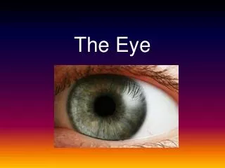
Outer Tunic • Made up of: - Cornea: clear avascular surface that allows light to enter eye - Sclera: protects the eye and is an attachment from extrinsic muscles - Optic Nerve: located at back of eye, a nerve that carries visual input to brain.
Middle Tunic • Made up of: - Choroid Coat: blood vessels here nourish surrounding tissues. Also contains melanocytes - Ciliary Body: thickest of middle tunic. Contains folds called ciliary processes. Helps with shape of pupil. Made up of ciliary muscles - Lens: Avascular. Made up of epithelial tissue. Behind iris and pupil - Iris: Colored portion of eye. Lies between cornea and lens • Amount of melanin distribution determines eye color - Pupil: Hole which light passes through. • Amount of light that enters is determined by size of opening of pupil
Inner Tunic • Made up of: - Macula Lutea: Yellowish spot. Provides central vision - Fovea Centralis: Depression in the center of the retina region. Produces sharpest vision - Optic Disc: Medial to fovea centralis. Nerve fibers from retina leave the eye to become part of optic nerve • Referred to as blind spot of the eye - Retina: • Photoreceptive sensory layer • Transparent sheet of continuous tissue • Transmit impulses • Contains neurons and nerve fibers • Neurons- bipolar cells, ganglion cells, amacrine cells, photoreceptor cells, horizontal cells
Neurons • Amacrine cells • Horizontal cells • Receptor cells • Ganglion cells • Bipolar cells
Amacrine Cells • Interneurons in the retina • Modify impulses on the fibers of the direct pathway • Help out horizontal cells
Horizontal Cells • Regulate input from photoreceptor cells which allow eyes to modify to light
Ganglion Cells • Located in inner surface of the retina • Received visual info. from bipolar cells & amacrine cells • Transmits image-forming visual info. from retina
Bipolar Cells • Transmits signals to the ganglion cells • Transmits signals to cones and rods
Receptor Cells • Consists of: - rods - cones
Cavities of the Eye • Anterior Cavity • Posterior Cavity
Anterior Cavity • Made up of: - Anterior Chamber: between cornea and iris - Posterior Chamber: between iris and vitreous humor • Filled with watery aqueous humor • Part of middle tunic
Posterior Cavity • Largest component of the eye • Filled with transparent, jellylike vitreous humor • Part of inner tunic
Accessory Organs • Eyelids: • Composed of 4 layers • Thinnest skin of the body • Open when: fibers of levator palpabrae superioris muscle arise from the roof of the orbit which then insert in the connective tissue of the upper lid • Contain sebaceous glands (tarsal glands)
Tarsal glands • sebaceous glands embedded in each eyelid • secrete oil along border of the lids • helps keep lids from sticking together
ACCESSORY ORGANS CONT. • Eyelashes: protect eyes from dust and dirt • Eyebrows: protects eyes from sweat or any chemical • Conjunctiva: Mucous membrane. Lines inner surface of eyelids. Covers anterior surface of eyeball’s lacrimal apparatus
Accessory Organs Cont. • Lacrimal Apparatus: - Lacrimal glands: secrete tears - 2 ducts: (superior and inferior canaliculi) which collect tears - Lacrimal sac: fluid from ducts move here - Nasolacrimal Duct: fluid from sac moves here and then empties in the nasal cavities
Accessory Organs cont. • Muscles:
Visual Receptors • Rods • Long thin projection at their terminal length • Sensitive to light • Produce outlines of objects • Produce colorless vision • Contains light-sensitive pigment: rhodopsin, or, visual purple
Cont. • Cones • Short blunt projections • Provide sharp images • Produce color images • Fovea centralis containing densely packed cones • Contains light-sensitive pigment: iodopsin
Refraction • Makes image formation possible • Light waves enter eye & an image of what is seen focuses upon retina • Light rays must bend to be focused; cornea & lens focus light • Light waves pass through the eye at an oblique angle • Convex lens- light comes together at one point • Concave lens- light waves are spread apart
Light goes through: • Cornea • Pupil • Lens • Vitreous Humor • Retina
Convergent v. Divergent • Divergent • Light focuses behind the retina • Convergent • Light focuses in front of retina
Accommodation • When eye focuses on an object • Lens thickens when focusing on a close object • Lens thins when object is distant
Stereoscopic Vision • Perceives distance, depth, height, and width of object • Depends on two eyed vision
RELATION TO BRAIN The occipital lobes of cerebrum are sensory areas responsible for vision Association areas combine visual images with other sensory experiences The midbrain of the brainstem contains reflex centers that move the eyes
NERVES SPECIFIC TO EYE & BRAIN Special somatic efferent fibers: carry sensory impulses inward to the brain from the receptors of sight. Found only in cranial nerves Cranial Nerves associated with eyes: Optic nerves (II): are sensory, and lead from the eyes to the brain and are associated with vision Oculomotor nerves (III): arise from midbrain and pass into orbits of eyes Trochlear nerves (IV): motor fibers transmit impulses to muscles that move the eyes
NERVES CONT. Trigeminal nerves (V): Ophthalmic Division: sensory fibers transmit impulses from surface of the eyes, tear glands, scalp, forehead, and eyelids Abducens nerves (VI): motor fibers transmit impulses to muscles that move the eyes Facial Nerves (VII): motor fibers transmit impulses to muscles of facial expression & tear glands
Bibliography • http://www.ivy-rose.co.uk/HumanBody/Eye/Anatomy_Eye.php • http://www.eyecareamerica.org/eyecare/anatomy/ • http://www.webmd.com/eye-health/picture-of-the-eyes • http://www.lensshopper.com/eye-anatomy.asp • http://hyperphysics.phy-astr.gsu.edu/hbase/vision/rfreye.html