Download
1 / 17
250 likes | 862 Vues
Pathogenic Mechanisms of Cancer Causing hMLH1 Mutations Functional Relationship between DNA Mismatch Repair and Tumor Formation. Eddie O’Donnell Laboratory of Dr. Andrew B. Buermeyer Department of Environmental and Molecular Toxicology. 2003 Estimated US Cancer Deaths. Men 285,900.

E N D
Pathogenic Mechanisms of Cancer Causing hMLH1 MutationsFunctional Relationship between DNA Mismatch Repair and Tumor Formation Eddie O’Donnell Laboratory of Dr. Andrew B. Buermeyer Department of Environmental and Molecular Toxicology
2003 Estimated US Cancer Deaths Men285,900 Women270,600 • 25% Lung & bronchus • 15% Breast • 11% Colon & rectum • 6% Pancreas • 5% Ovary • 4% Non-Hodgkin lymphoma • 4% Leukemia • 3% Uterine corpus • 2% Brain/ONS • 2% Multiple myeloma • 23% All other sites Lung & bronchus 31% Prostate 10% Colon & rectum 10% Pancreas 5% Non-Hodgkin 4%lymphoma Leukemia 4% Esophagus 4% Liver/intrahepatic 3%bile duct Urinary bladder 3% Kidney 3% All other sites 22% ONS=Other nervous system. *Excludes basal and squamous cell skin cancers and in situ carcinomas except urinary bladder. Source: American Cancer Society, 2003.
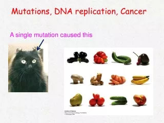
Colon Cancer • Causes of Cancer • Mutations within cells cause uncontrolled cell growth • Cancer may be inherited or sporadic • Colorectal Cancer • 15 % - Mismatch Repair (MMR) deficiency observed • 90 % of Sporadic cases linked to MMR deficiency are mlh1 • deficient (loss of expression) • 2-5 % - Hereditary Non-Polyposis Colorectal Cancer (HNPCC) • Recent Discoveries involving HNPCC • 1993 – MSH2 mutationslinked to 35% of HNPCC • 1994 – hMHL1 mutations linked to 35 % of HNPCC HNPCC, FAP Non Hereditary
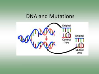
G T G T T A T DNA Mismatch Repair • DNA Mismatches arise from errors during DNA Replication • MMR is an essential process for maintaining genomic integrity • Basic Mechanism: • Mismatch recognition * • Strand choice • Excision • Resynthesis
Mutation Types • DNA Synthesis Error Mutation • Base Mismatches Base Substitution Mutations • Insertion / Deletion Loops Microsatellite Instability (MSI) • Microsatellite loci are common and unique to a persons genome • Looking specifically at dinucleotide repeated sequences • Deficient MMR can lead to increased probability of replication errors No Repair, Continued Replication Incorrect DNA sequence, mutation in genome copy Incorrect Placement of Base A T A T G T Successful Repair G C Incorrect DNA sequence, mutation in genome copy Dinucleotide Loop Insertion No Repair, Continued Replication AC TG Successful Repair
Clinical relevance of the hMLH1 gene • hMLH1 significance • hMLH1 is an essential protein for the prevention of • mutations. Exact function is unknown. • Treatments • MMR deficient cancers may respond differently • to chemotherapeutic drugs. • Detection • MMR deficient cancers are commonly detected • through screening for MSI, however… • Several hMLH1 Mutations have been implicated in HNPCC cases showing only high levels of base substitution mutations: not initially identified as MMR deficient cancers. E578G K618A D132H V716M hMLH1 amino acid site 578 changed from E (glutamic acid) to G (Glycine)
Preliminary Research with hMLH1 E578G • Data from analysis of cells expressing E578G demonstrate: • No MSI, consistent with observed cancers • Increased levels of base substitution mutations • E578G mutation affects repair of base base • mismatches but not dinucleotide loops • Increased base substitutions may explain pathogenicity of mutations • suggests a possible novel role for MMR repair genes, containing • MLH1, in substrate recognition and / or commitment to initiate repair
Research Questions & Goals • Are mutations of hMLH1 responsible for the observed molecular phenotypes? The goal of the research is to determine the mutation prevention capabilities of the MLH1 mutants E578G, K618A, V716M, and D132H using biochemical assays
Project Overview Generation of substrates to model DNA mismatches Stable transfections create extracts containing repair factors including mutant hMLH1 protein Biochemical Assays will elucidate the repair efficiency of the mutant hMLH1 protein
Biochemical Assay Procedure Length of repair products will be measured with Analytical gel electrophoresis.
Selected Cell Lines Positive Control Negative Control Cytoplasmic Extracts Stable Transfection • Mutant hMLH1 gene introduced via electroporation • A specific sequence , unrelated to the hMLH1 gene, which enables resistance to the drug G418 is also introduced to allow for selection of cells that were succesfuly transfected • Cells expressing the desired MLH1 mutation provide the source for MLH1 mutant protein and other repair proteins • Selection of cell lines expressing sufficient levels of MLH1 mutants relative to wildtype MHL1 using western blotting Extracts E578G Cell Line Selection 250 kD 150 kD MSH6 (140.1 kD) 100 kD PMS2 75 kD MLH1 ß-Tubulin (50.9 kD) 50 kD
Substrate Formation • Research will involve in-vitro MMR reactions and different plasmid • substrates, that model presumed replication errors, with either • -single base pair mismatches model base substitutions • -dinucleotide insertion loops model MSI mutations Contaminating Plasmid T G - + Purified Substrate
Substrate Formation A circular piece of DNA, known as a plasmid, serves as the starting material for the mismatch repair substrate. A nicking enzyme, which cuts one strand of a double stranded sequence, cuts at two sites on the plasmid DNA. The Plasmid is heated to remove the cut fragment, and the complementary strand is added to create a pure gap molecule A DNA oligo, similar to removed fragment, is introduced to the gap molecule, and DNA Ligase is used to anneal the mismatch oligo to form the mismatch substrate.
Substrate Formation Diagnostics used at each step to ensure quality of substrate • Does the starting plasmid cut correctly? • How efficient was the nicking of the DNA • Purity of gap molecule ? • Is the gap molecule resistant to cutting ? • Is the mismatch substrate resistant to cutting ? Gap Molecule Mismatch Substrate Nicked Plasmid Starting Plasmid Nicked DNA Linearized DNA Supercoiled DNA Linear DNA from double digest Uncut Plasmid Restriction Endonuclease Cut
Preliminary Repair Assays • Cellular extracts without MLH1 protein and extract with functional (wildtype) MLH1 • protein will serve as experimental controls Test Repair Assays Extracts: A – HeLa cells (positive control) B – WT22 cells (positive control) C – E578G D – CMV2 (negative control) E – No extract (negative control)
Further Research • Questions • - Do any of the MLH1 mutants show ability to differentiate • Between different types of mismatches • At what efficiency relative to wildtype MLH1 do the • mutants repair mismatches
Thank You • HHMI • URISC • Dr. Andrew Buermeyer • The Buermeyer Lab Group. “Good People” • Dr. Kevin Ahern