Download
1 / 13
130 likes | 273 Vues
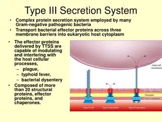
The Motion of a Single Molecule, the λ-Receptor, in the Bacterial Outer Membrane Oddershede, Dreyer, Grego, Brown, and Berg-Sørensen Biophysical Journal Volume 83 December 2002. staphylococci streptococci. K. pneumoniae E. coli. The λ-Receptor. Biotinylation site. ▼.

E N D
The Motion of a Single Molecule, the λ-Receptor, in the Bacterial Outer Membrane Oddershede, Dreyer, Grego, Brown, and Berg-Sørensen Biophysical Journal Volume 83 December 2002
staphylococci streptococci K. pneumoniae E. coli
The λ-Receptor Biotinylation site ▼
Purpose of the model To extract a series of parameters describing the biological system • γp : friction the protein feels in the membrane • D : diffusion coefficient of the protein passing through the membrane kBT ____ D = γp
κ : spring constant of bead in optical tweezers • κbs : spring constant of attachment to bead • κ cw : spring constant of attachment to cell wall • χcw : position of protein and cell wall • χp : position of protein • χb : position of the bead • χtrap : position of optical trap
/ Streptavidin - bead Receptor-biotin \ Streptavidin - FITC
Results Low biotinylation rate
FIGURE 2 Position of a bead on λ-receptor versus time FIGURE 3 Position histogram of a bead on -receptor (squares) and of a bead unattached to a bacterium (circles) found by optical tweezers.
FIGURE 4 Power spectrum of x(t) for a bead on λ-receptor (referred to as bacterium) and for an unattached bead (reference) held by optical tweezers.
FIGURE 6 Data from a bead attached to -receptor in the bacterial membrane obtained by SPT (a) The x-position of the bead (b) Histogram of the position distribution
FIGURE 6 Data from a bead attached to -receptor in the bacterial membrane obtained by SPT (c) Scatter plot, showing the distribution of locations of the bead within the plane of the bacterial membrane. (d) Mean square displacement (MSD) as a function of time taken over 50 averages.
Conclusions Method for in vivo biotinlytion of the λ-receptor, but can also be used for protein motion in other membrane systems γp and D are independent of laser intensity κ cw : 1.1 x 10 –2 pN/nm by OT κ cw : 0.98 x 10 –2 pN/nm by SPT D = 1.5 x 10⁻⁹ cm2/s Motion of the λ-receptor is energy dependent – to facilitate transport of maltodextrin? } identical