Ch. 10 Blood
200 likes | 356 Vues
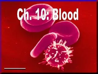
Ch. 10 Blood. The only fluid tissue in the human body Average body has 5 to 6 liters or 6 quarts Classified as a connective tissue Living cells = formed elements Non-living matrix = plasma Composition Plasma – 55% of blood is plasma ; clear watery part of blood

Ch. 10 Blood
E N D
Presentation Transcript
Ch. 10 Blood • The only fluid tissue in the human body • Average body has 5 to 6 liters or 6 quarts • Classified as a connective tissue • Living cells = formed elements • Non-living matrix = plasma • Composition • Plasma – 55% of blood is plasma ; clear watery part of blood • Made of 90% of water; used to carry other substances which acts as a solvent • Salts – allows for the proper osmotic balance and pH buffering • Plasma proteins – clotting of blood, defense (antibody); made mostly by the virus
Formed Elements – living blood cells • Erythrocytes - RBC; boat for cells needed to be carried to the body • Leukocytes – WBC; used to fight off bacteria, viruses, parasites & tumor cells (foreign bodies) • Platelets – not cells; used to help clot blood
Plasma • Composed of approximately 90 percent water; used to carry other substances which acts as a solvent • Includes many dissolved substances • Nutrients • Salts (metal ions) – allows for proper osmotic balance & pH buffering • Respiratory gases • Hormones • Proteins • Waste products • Plasma Proteins • Albumin – regulates osmotic pressure • Clotting proteins – help to stem blood loss when a blood vessel is injured • Antibodies – help protect the body from antigens
Formed Elements • Erythrocytes = red blood cells • Leukocytes = white blood cells • Platelets = cell fragments
Erythrocytes – RBC • The main function is to carry oxygen • Anatomy of circulating erythrocytes • Biconcave disks • Essentially bags of hemoglobin • Anucleate (no nucleus) • Contain very few organelles • Outnumber white blood cells 1000:1 • Hemoglobin • Iron-containing protein • Binds strongly, but reversibly, to oxygen • Each hemoglobin molecule has four oxygen binding sites • Each erythrocyte has 250 million hemoglobin molecules • Anemia – disorder causing decreasing amounts of O2 being carried to the bodies because of either • Low numbers of RBC • Abnormal shapes causing less O2 preventing proper carrying
Leukocytes – WBC • Crucial in the body’s defense against disease • These are complete cells, with a nucleus and organelles • Able to move into and out of blood vessels (diapedesis) • Can move by ameboid motion • Can respond to chemicals released by damaged tissues • Normal levels are between 4,000 and 11,000 cells per millimeter • Abnormal leukocyte levels • Leukocytosis • Above 11,000 leukocytes/ml • Generally indicates an infection • Leukopenia • Abnormally low leukocyte level • Commonly caused by certain drugs
Types of Leukocytes • Granulocytes • Granules in their cytoplasm can be stained • Include neutrophils, eosinophils, and basophils • Agranulocytes • Lack visible cytoplasmic granules • Include lymphocytes and monocytes
Granulocytes • Neutrophils • Multilobed nucleus with fine granules • Act as phagocytes at active sites of infection • Eosinophils • Large brick-red cytoplasmic granules • Found in repsonse to allergies and parasitic worms • Basophils • Have histamine-containing granules • Initiate inflammation • Agranulocytes • Lymphocytes • Nucleus fills most of the cell • Play an important role in the immune response • Monocytes • Largest of the white blood cells • Function as macrophages • Important in fighting chronic infection
Platelets • Derived from ruptured multinucleate cells (megakaryocytes) • Needed for the clotting process • Normal platelet count = 300,000/mm3 • Hematopoiesis - blood cell formation • RBC live for only 100 to 120 days • The spleen & liver produces phagocyctes which destroy decaying RBC • Occurs in red bone marrow • All blood cells are derived from a common stem cell (hemocytoblast) • Hemocytoblast differentiation • Lymphoid stem cell produces lymphocytes • Myeloid stem cell produces other formed elements
Hemostasis • Stoppage of blood flow • Result of a break in a blood vessel • Hemostasis involves three phases • Platelet plug formation • Vascular spasms • Coagulation • Platelet plug formation • Collagen fibers are exposed by a break in a blood vessel • Platelets become “sticky” and cling to fibers • Anchored platelets release chemicals to attract more platelets • Platelets pile up to form a platelet plug • Vascular spasms • Anchored platelets release serotonin • Serotonin causes blood vessel muscles to spasm • Spasms narrow the blood vessel, decreasing blood loss
Coagulation • Injured tissues release thromboplastin • PF3 (a phospholipid) interacts with thromboplastin, blood protein clotting factors, and calcium ions to trigger a clotting cascade • Prothrombin activator converts prothrombin to thrombin (an enzyme) • Thrombin joins fibrinogen proteins into hair-like fibrin • Fibrin forms a meshwork (the basis for a clot) • Blood Clotting • Blood usually clots within 3 to 6 minutes • The clot remains as endothelium regenerates • The clot is broken down after tissue repair
Undesirable Clotting • Thrombus • A clot in an unbroken blood vessel • Can be deadly in areas like the heart • Embolus • A thrombus that breaks away and floats freely in the bloodstream • Can later clog vessels in critical areas such as the brain • Bleeding Disorders • Thrombocytopenia • Platelet deficiency • Even normal movements can cause bleeding from small blood vessels that require platelets for clotting • Symptoms – small purple blotches on skin; cure vitamin K • Hemophilia • Hereditary bleeding disorder • Normal clotting factors are missing • Symptoms – prolong bleeding; cure injection of clotting factors
Blood Groups & Transfusions • Large losses of blood have serious consequences • Loss of 15 to 30 percent causes weakness • Loss of over 30 percent causes shock, which can be fatal • Transfusions are the only way to replace blood quickly • Transfused blood must be of the same blood group • Human Blood Group • Blood contains genetically determined proteins • A foreign protein (antigen) may be attacked by the immune system • Blood is “typed” by using antibodies that will cause blood with certain proteins to clump (agglutination) • There are over 30 common red blood cell antigens • The most vigorous transfusion reactions are caused by ABO and Rh blood group antigens
ABO Blood Groups • Based on the presence or absence of two antigens • Type A • Type B • The lack of these antigens is called type O • The presence of both A and B is called type AB • The presence of either A or B is called types A and B, respectively • The wrong blood type can cause the bodies natural defense (antibodies) to clot; could also cause fever, chills, nausea & vomiting
Rh Blood Groups • Named because of the presence or absence of one of eight Rh antigens (agglutinogen D) • Most Americans are Rh+ • Problems can occur in mixing Rh+ blood into a body with Rh– blood
Rh Dangers During Pregnancy • Danger is only when the mother is Rh– and the father is Rh+, and the child inherits the Rh+ factor • The mismatch of an Rh– mother carrying an Rh+ baby can cause problems for the unborn child • The first pregnancy usually proceeds without problems • The immune system is sensitized after the first pregnancy • In a second pregnancy, the mother’s immune system produces antibodies to attack the Rh+ blood (hemolytic disease of the newborn)
Blood Types • Blood samples are mixed with anti-A and anti-B serum • Coagulation or no coagulation leads to determining blood type • Typing for ABO and Rh factors is done in the same manner • Cross matching – testing for agglutination of donor RBCs by the recipient’s serum, and vice versa