Download
1 / 57
570 likes | 712 Vues
This comprehensive overview explores the syndromes of stroke and head trauma, focusing on rapid onset cerebral deficits. It discusses risk factors, differentiating between non-modifiable and modifiable elements. Various types of strokes, including ischemic and hemorrhagic, are examined alongside their causes. The document outlines key symptoms, assessment techniques, acute interventions, and the critical role of diagnosis through imaging. Additionally, it touches on complications and recovery challenges, emphasizing the need for immediate treatment and secondary prevention strategies.

E N D
Stroke, Head Trauma and conciousness Amy Wood, Haddy Cosh, Vishal Chauhan, AsfandBaig, Stewart O’Conner
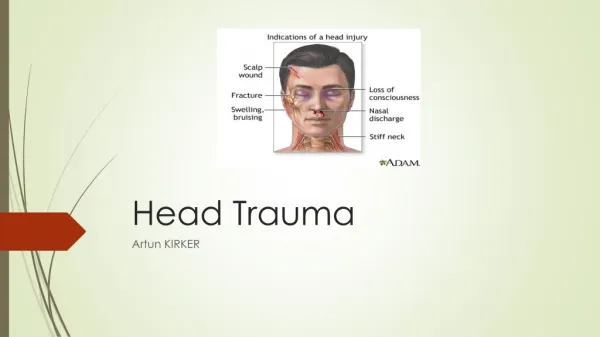
Definition • a syndrome of rapid onset of cerebral deficit (usually focal) • Lasting > 24 hours or leading to death and no cause apparent other than a vascular one
Stroke Risk Factors • Non Modifiable • Modifiable
Stroke Risk Factors • Non Modifiable • Age • Male • FHx • Race – black/ hispanic • Modifiable • HT • IHD • AF • DM • Hypercholesterolaemia • Smoking • Alcohol
Types • Ischaemia/ embolism causing cerebral infarct – 80% • Intracebral Haemorrhagic – 15%
Causes -Haemorrhagic • Ruptured aneurysm • Trauma (subarachnoid/intracerebral) • Dissection (carotid/vertebral)
Causes - Ischaemic • Cerebral Thrombosis • Cerebral Emboli • Give examples • Lacunar
Symptoms - General • Weakness/Paralysis or numbness on contralateral side • Vertigo/dizziness • Headache • Visual loss/blurred vision • Faintness • Confusion • Speech problems • Difficulty swallowing • Cognitive problems • Memory problems • Consciousness alterations • BUT…DEPENDS ON SITE
Stroke Syndromes • TACS - Total Anterior Circulation Syndrome • PACS - Partial Anterior Circulation Syndrome • LACS - Lacunar Syndrome • POCS - Posterior Circulation Syndrome
Extras - watersheds • Susceptibility to ischaemia: • Systemic BP drop • ACA-MCA occlusion of carotid
TIA • Sudden focal deficit – usually only a few seconds • Presentation very similar to stroke • Amaurosis fugax?? • <24 hours with complete recovery • Issue: after 1 hour ischaemic damage has already occurred • High risk of recurrence and full stroke
Causes- TIA Carotid artery insufficiency – 80% Veterbrobasilar Insufficiency – 20% Circle of Willis – collateral supplies
Management • Assessment/ diagnosis • Location • Subtype • Cause • Acute intervention • Secondary prevention • Reduce risk factors
Assessment: Diagnosis • Clinically usually • FAST • Imaging - <3hrs • CT • Available • Exclude haemorrhage • MRI • If brainstem or cerebellar symptoms
Acute intervention • Admit to Acute Stroke Unit for assessment • Iscahaemic – Thrombolysis rTPA within 3 hrs of symptoms • Haemorragic – emergency surgery
Acute intervention • Antiplatelet drugs (Aspirin 150-300mg) if infarct • Contraindicated if haemorrhage!! • Monitor/prevent complications • Physiological monitoring for first 72 hours to maintain CO and supply to brain • HR, Temperature, BP, O2 sats, Blood sugar, ECG
Complications • Post-stroke pain/thalamic pain • 1 week- 6 months after stroke • Anywhere in spinothalamic system • Contralateral side referral of pain • Burning + sharp • Hyperalgesia & Allodynia • Treat as for neuropathic pain • TCAs
Layers of the brain a) Pia mater b) Arachnoid mater c) Dura mater d) Superior sagittal venous sinus e) Skull f) Falxcelebri g) Subarachnoid space
Pia • Arachnoid • Dura Subarachnoid – arteries Subdural – Bridging veins Epidural – Meningeal arteries
Normal CT • Usually going to be symmetrical • Ventricles symmetrical and equally full
Midline Shift • Coup injury – injury on same side of force • Contra coup– injury on the opposite side on injury • If you see midline shift, you have a high pressure situation
Case 1 • Young lady hit on the side of head by a glass at a gig, seemed to recover , Found slumped 50 minutes later • Ix? • CT/MRI, x-ray if fracture • Where may she have been hit? • Pterion • What bones converge here? • frontal, parietal, sphenoid, temporal • What does this area cover? • Middle meningeal artery • Type of intracranial haemorrhage? • extradural (epi) • Type of blood characterises this? • Arterial • Why passed out? • raised ICP • Rx • surgical
Extraduralhaematoma: • Midline shift • Lenticular shape • This can be middle meningeal artery – pterion bone breaks • Cerebral perfusion pressure = mean arterial pressure – ICP • Extraduralhaematoma you give Mannitol – 100mL at 20% • Diuretic
Case 2 • Old alcoholic man had a fall in the park now noticed to be very drowsy with low consciousness • Ix: • CT/MRI • Likely haematoma? • Subdural • Other symptoms? • Headache, confusion, N/V, tinnitus, speech and visual problems, dizziness, weakness • Where is the bleed likely to be? • bridging veins • Type of blood? • venous • Rx depends on size + growth rate: often conservative (body reabsorbs), sometimes burr-hole drainage • Acute or Chronic
Subdural Haematoma: • Runs along the surface of the brain, underneath the dura • Depending on the GCS score of the patient you may need to remove it • Midline shift
Subarachnoid Haemorrhage • Sudden onset severe headache, often at the back of the head, Neck stiffness, Impaired consciousness (drowsiness / coma), Cranial nerve signs, Hemiplegia • The bleeding occurs as the result of rupture of aneurysm (80%) and AV malformations (15%) or trauma
Contusion (bruise) • Intra- axial • As bruise swells, pressure goes up – all features of raised ICP (coma) • If you remove them you need to do a craniotomy
Diffuse Axonal InjuryRTAs / shaken baby syndrome • If a rotational force is applied, the axons are damaged and you can have damage very far away from the original injury – diffuse axonal injury • Small contusions all over the brain • The worse it looks on the CT scan, the worse the injury in the patient – especially if you see an injury in the brainstem • DAI doesn’t look as bad on CT as some of the other ones, but can be much worse
With a mass lesion why do you not get an immediate loss of consciousness?
Due to an ability to Compensate! Intra cranial vol = vol CSF + vol Brain + vol blood + vol Mass lesion Skull can’t expand Compensation – 10-20 ml CSF in to lumbar cisterns Compensation exceeded Increase in ICP herniation
What are the 3 key symptoms of raised ICP? Papilloedema Headache Nausea and Vomiting