Download
1 / 43
430 likes | 517 Vues
Learn about the cardiovascular system, the heart's functions, how it works, and the circulation of blood through the body. Understand the roles of arteries, capillaries, and veins in maintaining circulation.

E N D
CHAPTER 3 - CIRCULATIONChapter 1: Section 1 – The Body’s Transport System Mrs. Swanson
I. The Cardiovascular System • A.Circulatory system1.Heart, blood vessels, and blood2.Body’s transportation systemB.Three major functions: 1.delivering needed materials 2.removing waste products 3.fighting disease
II. The Heart - a hollow, muscular organ that pumps blood throughout the body • A.Facts:· About the size of your fist· Located in your chest behind your sternum · Each time it the heart beats, it pushes blood through three blood vessels· Composed of cardiac muscle
B. Structure (use diagrams) • 1. Two sides (right and left based on person’s perspective)2. Separated by the septum (wall of tissue)3. Atrium (upper chamber)- receives blood that comes into the heart4. Ventricle (lower chamber)- pumps blood out of the heart5. Atria and ventricles are separated by valves- a flap of tissue that prevents blood from flowing backward (like a one-way door).
Web Sites • http://www.kidshealth.org/kid/body/heart_noSW.html • http://www.phschool.com/webcodes10/index.cfm?wcprefix=cep&wcsuffix=4031&fuseaction=home.gotoWebCode&x=0&y=10
C. How the Heart Works • 1.The heart has two main phases-2.Heart muscle relaxes - atria fills with blood 3.Heart muscle contracts - pumps blood through the valves and into the ventricles.4.Ventricles contract, closing the valves between the atria and the ventricles (lub sound)5.The ventricles - (dub sound) blood enter the arteries.6.Right ventricle -blood to the lungs 7.Left ventricle- blood throughout body
D. Regulation of Heart Beat • 1.Pacemaker – a group of heart cells which sends out signals that make the heart muscle contract (located in the right atrium). • 2.When a person’s pacemaker has been damaged, it can cause slow or cause irregular heartbeats can be replaced with artificial pacemakers
Journal • Draw a quick Heart and describe the movement of blood through the heart and to the lungs and body. • Hint: Start in the Right Atrium
E. Vessels • 1. Three types of blood vesselsa.Arteries - blood vessels that carry blood away from the heartb.Capillaries –narrow vessels receive blood from the arteries, exchange O2 and CO2 between the blood and body cellsc.Veins - blood vessels carry blood back to the heart.2. Blood travels in one direction: • Heart, Lungs, Heart, Body, Heart
Loop One: To the Lungs and Back • · Blood entering right atrium has little oxygen and a lot of carbon dioxide (waste product) - This oxygen-poor blood is dark red (not blue like seen in pictures)· Blood flows from right atrium into right ventricle· Blood flows from the right ventricle into the arteries that lead to lungs (to get more oxygen)· Arteries branch into smaller capillaries inside the lungs so that oxygen can move into the blood· Oxygen-rich blood (bright red) leaves the lungs and flows to the left side of the heart to enter into the second loop
Loop Two: To the Body and Back • ·Left atrium fills with oxygen-rich blood from the lung· Blood flows from the left atrium in to the left ventricle · From the left ventricle, the blood is pumped into the aorta (largest artery in to body)· Blood flows through large arteries which branch into smaller capillaries inside different body parts-Oxygen moves out of the blood and into the body cells-Carbon dioxide moves out of the body cells and into the blood· Oxygen-poor blood flows back to the right atrium (this completes loop two where it will begin again with loop one
Chapter 1: Section 2 – A Closer Look at Blood Vessels • Blood vessels are like hallways in a building – they run throughout your entire body· Can be as wide as your thumb; most are as thin as a tiny hair· All blood vessels hooked together end to end would stretch a distance of almost 100,000 kilometers (more than 2x around the earth)
I. Arteries- Transports blood AWAY FROM the heart • A. Artery Structure 1.Walls are generally very thick- three cell layers give strength and flexibility.a. Epithelial Tissue layer– innermost; smooth to enable easy blood flowb. Muscle Tissue layer– middle; gives strengthc. Connective Tissue layer– outermost; gives flexibilityB. Pulse- the alternating expansion and relaxation of the artery wall.
ARTERIES • http://www.phschool.com/atschool/phsciexp/2005/human_biology_and_health.html#
C. Regulating Blood Flow • 1.The muscle tissue layer - the control gate, adjusts the amount of blood sent to different organs2.When the muscle contracts, the artery becomes smaller (less blood flow)3.When the muscle relaxes, the artery becomes larger (more blood flow)4.E.g. – after you eat, your digestive organs need more blood to go through digestion, so the arteries will open wider allowing for more blood flow
II. Capillaries- these cells are only one layer thick (epithelial cells). • A. Structure- • 1.Oxygen and glucose pass out of blood to cells; carbon dioxide passes out of cells into blood by means of diffusion.2.Diffusion –molecules move from an area of higher concentration to an area of lower concentration
III. Veins • A. Structure- walls have three layers (inner epithelial, middle muscle, outer connective), thinner than the arteries.2.The heart does not move the blood in the veins (because veins are farther away from the heart). Several factors do • a.skeletal muscles - contract to help push bloodb.valves- prevent blood from flowing backwardc.Diaphragm- and lungs- exert a squeezing pressure on the veins
IV. Blood Pressure- the force blood exerts against the walls of blood vessels (ventricles) • A. Measuring Blood Pressure- sphygmomanometer (sfig moh muh NAHM uh tur)= blood pressure cuff.1. Measured in millimeters of mercury3.Records two numbersa.First number–pressure of blood leaving ventricles and entering arteriesb.Second number - while the ventricles relax (this number is lower)c.Expressed in a fraction: contraction pressure/relaxation pressure
Heart Beat/ Health Beat • Pg. 90 in textbook. • Data Share when you are done • www.phschool.com • Web code ced-4032
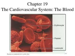
Chapter 3: Section 3 - Blood and Lymph • I. BloodA.blood is made up of four components: plasma, red blood cells, white blood cells, and platelets.B.about 45% of blood volume is cells, 55% is plasma
Components White Blood Cell Red Blood Cell Plasma Cell
II. Plasma- liquid part of the blood • A.90% of plasma is water; 10% is dissolved materials (glucose, fats, vitamins & minerals) carry away waste, yellow in color. • 1. Three groups of plasma proteins:a.regulate the amount of water in bloodb. (made by white blood cells) helps fight diseasec.interacts with platelets to form blood clots
II. Red Blood Cells • A.Function- take up oxygen in lungs and bring it to everywhere in the body1.produced in bone marrow2.disk-like shape with pinched-in center allows for easy movement through narrow blood vessels3.made mostly of hemoglobin (HEE muh gloh bin)-iron- containing protein that binds chemically to oxygen molecules making it BRIGHT REDB.Life Span- no nuclei, can not reproduce 1.live for about 120 days2.every second about 2 million die
III. White Blood Cells • A.Function1. help the body fight disease2. Alert the body when invaded by disease 3. Produce chemicals to fight the invaders4. Surround and kill invaders
IV. Platelets-cell fragments forming blood clots • A. Steps of blood clot formation:1.Blood vessel is cut2.Platelets collect and stick to vessel at site of wound3. Platelets produce fibrin (weaves a net of tiny fibers)4.Fiber net traps blood vessels and platelets forms a blood clot (a scab)
V. Blood Types • A. Blood transfusion – transfer of blood from one person to another1.Early blood transfusions failed until the early 1900s when Karl Landsteiner tried mixing blood samples from pairs of people.2.Sometimes the samples blended smoothly, other times they clumped preventing a successful blood transfusion
B. Marker Molecules- Landsteiner discovered 4 major types of blood - A, B, AB, O • 1. Blood types are determined by proteins known as marker molecules on red blood cells.a. A blood type - A markersb.B blood type –B markersc.AB blood type – both A and B markersd.O blood type – neither A nor B markers2. Plasma contains clumping proteins that recognize “foreign” markers and make those cells clump togetherE.g. – If you have A blood type, and get a transfusion of B blood, your clumping proteins will make the foreign” type B cells clump together
Marker Molecules in the Blood • 4.The marker molecules on your red blood cells determine your blood type and the type of blood that you can safely receive in transfusions • 5.Examples of safe transfusions:
Blood Typing • http://nobelprize.org/educational_games/medicine/landsteiner/readmore.html
The Lymphatic System • · Lymphatic system– a network of vein-like vessels that returns the fluid to the blood stream· Carries excess fluid away • Exchange at the capillaries
Lymph • · lymph-water, dissolved materials such as glucose, and white blood cells that have left the capillaries • · moves slowly (not being pumped)· connect to veins in chest where the lymph empties into the blood plasma
Lymph Nodes • · Within lymphatic vessels there are small knobs of tissue called lymph nodes· Filter lymph (trapping bacteria and other disease-causing microorganisms)· When the body is fighting an infection, lymph nodes enlarge (swollen glands)
Chapter 3: Section 4 - Cardiovascular HealthCardiovascular DiseasesLeading cause of death in the United States today • ·Atherosclerosis (ath uh roh skluh ROH sis) – a condition in which an artery wall thickens as a result of the build up of fatty materials (such as cholesterol)· Results in a reduced flow of blood· Can develop in the coronary arteries, which could lead to reduced blood flow and oxygen to the heart (may cause heart attack)· Heart attack - occurs when blood flow to part of the heart muscle is blocked; cells die here; permanently damages the cell of the heart·
Types of treatment: • a. low-fat diet (mild atherosclerosis) b. moderate exercise program (mild atherosclerosis)c. medications that lower cholesterol and fats in blood (mild atherosclerosis)d.surgery or other procedures to unclog blocked arteries (severe atherosclerosis