Download
1 / 51
510 likes | 679 Vues
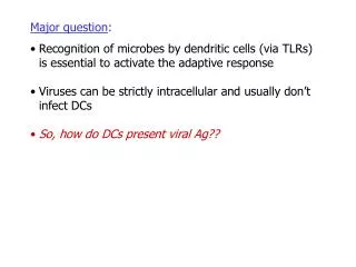
Major question : Recognition of microbes by dendritic cells (via TLRs) is essential to activate the adaptive response Viruses can be strictly intracellular and usually don’t infect DCs So, how do DCs present viral Ag??. Hypotheses :. Hypotheses :

E N D
Major question: • Recognition of microbes by dendritic cells (via TLRs) is essential to activate the adaptive response • Viruses can be strictly intracellular and usually don’t infect DCs • So, how do DCs present viral Ag??
Hypotheses: • Cells other than DCs (stromal cells) contribute to presentation of Ag • TLRs of stromal cells recognize pathogen-associated molecular patterns (PAMPs)
Experimental system: • Herpes simplex virus type 2 (HSV-2) • Thymidine kinase (TK) mutant can infect but not replicate • Infect vaginal epithelium of mice • Examine response to mucosal infection • Primarily measure activation of CD4+ TH1
Figure 1: IF-THEN??
Figure 1: IF TLR signalling is required for TH1 response to HSV-2, THEN disrupting TLR signalling will block TH1 response
Figure 1: Technique for measuring TH1 response?
Figure 1: • Technique for measuring TH1 response: • Infect mice with HSV-2 • Isolate CD4+ cells from lymph nodes • anti-CD4 on magnetic beads • Stimulate in vitro with APCs or DCs ± HSV-2 Ag • Assay cytokine production • how?
Figure 1: • Disruption of TLR signalling: • MyD88 is an adapter protein in the TLR signal transduction pathway • Knockout mice (MyD88 –/–) lack this protein (why is this better than knocking out a TLR?)
Figure 1: • Activated cells in WT make IFN-g, not IL-4 or 10 • Activated cells in KO make IL-4 and 10, not IFN-g APCs DCs Significance? Controls?
Figure 1: • Activated cells in WT make IFN-g, not IL-4 or 10 • Activated cells in KO make IL-4 and 10, not IFN-g • TLR signalling is needed for TH1 response APCs DCs
Figure 2: IF-THEN??
Figure 2: IF TLR signalling is required for DC recruitment to mucosa, THEN disrupting TLR signalling will block recruitment
Figure 2: Technique for measuring recruitment to mucosa?
Figure 2: • Technique for measuring recruitment to mucosa: • Immunofluorescence staining (in situ) • anti-CD11c Ab: specific DC marker
Figure 2: • Similar recruitment of DCs to infected epithelium WT KO blue = nuclei red = DCs green = virus
Figure 3: IF-THEN??
Figure 3: IF TLR signalling is required for DC migration to lymph nodes or maturation, THEN disrupting TLR signalling will block migration or maturation
Figure 3: Technique for measuring migration to lymph nodes and maturation?
Figure 3: • Technique for measuring migration to lymph nodes and maturation: • Fluorescence-activated cell sorting (FACS)
Figure 3: • Technique for measuring migration to lymph nodes and maturation: • Fluorescence-activated cell sorting (FACS)
Figure 3: • Technique for measuring migration to lymph nodes and maturation: • Fluorescence-activated cell sorting (FACS) • DCs are: CDC11c+ / B220– / CD8– • Mature DCs are also CD86+ • Isolate CD11c+ cells • Sort with fluorescent anti-CD8 and anti-CD86
Figure 3: • TLR signalling does not change numbers of CD4 or DC cells in lymph nodes
Figure 3: • TLR signalling does not change numbers of CD4 or DC cells in lymph nodes • TLR signalling does not change number or maturity of DCs Mock HSV WT KO mature DCs
Figure 3: • TLR signalling does not change numbers of CD4 or DC cells in lymph nodes • TLR signalling does not change number or maturity of DCs • TLR signalling is not needed for migration or maturity Mock HSV WT KO mature DCs
Figure 4: • Possible explanations for Fig. 1 results: • TH1 activation requires TLR signalling • TH1 activation requires IL-1 or IL-18 (also dependent on MyD88) • TH1 activation requires IL-12 • TH1 activation requires IFN-g
Figure 4: IF-THEN??
Figure 4: IF MyD88 KO mice lack TH1 response due to lack of IL-1 (or IL-18), IL-12 or IFN-g, THEN lack of one of these products would have the same effect as the MyD88 KO
Figure 4: Technique for measuring effects of cytokines on TH1 activation?
Figure 4: • Technique for measuring effects of cytokines on TH1 activation: • Additional KO mice: • caspase-1 (IL-1 b-converting enzyme) KO mice: lack functional IL-1 • IL-12 p40 KO mice: lack functional IL-12 • IFN-gR KO mice: DCs can’t respond to IFN-g • Same cytokine assay as Fig. 1
Figure 4: • Lack of IL-1 does not affect TH1 activation • Lack of IL-12 does not affect TH1 activation • Lack of DC response to IFN-g does not affect TH1 activation APCs DCs
Figure 4: • Lack of IL-1 does not affect TH1 activation • Lack of IL-12 does not affect TH1 activation • Lack of DC response to IFN-g does not affect TH1 activation • Activation appears to specifically require TLR signal APCs DCs
Figure 5A: IF-THEN??
Figure 5A: IF epithelial cells are involved in TLR signalling, THEN these cells must express TLR genes
Figure 5A: Technique for measuring expression of TLR genes?
Figure 5A: • Technique for measuring expression of TLR genes: • RT-PCR • Primer pair specific for each TLR gene • Isolate mRNA • Reverse transcriptase DNA • PCR to amplify: band indicates mRNA was present
Figure 5A: • Epithelial cells express all tested TLR genes
Figure 5B-E: IF-THEN??
Figure 5B-E: IF TLR signalling by epithelial cells is needed for TH1 activation THEN MyD88 KO in epithelial cells will block TH1 activation even if WT DCs are present
Figure 5B-E: Technique for making mice with genetically different epithelial and DC cells???
Figure 5B-E: • Technique for making mice with genetically different epithelial and DC cells: • Bone marrow (BM) chimera: • lethal irradiation of mouse to kill bone marrow cells • “bone marrow transplant” from a different strain • epithelial cells have original genotype; DCs have donor genotype MyD88 (-/-) DCs MyD88+ epithelium WT MyD88 KO BM cells KO BM WT
Figure 5B-E: • Technique for making mice with genetically different epithelial and DC cells: • Bone marrow (BM) chimera • Same cytokine assay for CD4 cell activation
APCs DCs • Figure 5B-E: • MyD88 required in DCs for activation MyD88 (-/-) DCs MyD88+ epithelium
APCs DCs • Figure 5B-E: • MyD88 required in epithelial cells for activation MyD88 (-/-) epithelium MyD88+ DCs
APCs DCs • Figure 5B-E: • MyD88 required in DCs for activation • MyD88 required in epithelial cells for activation • Both DCs and stromal cells participate in Ag presentation and require TLR signalling
Figure 6: IF-THEN??
Figure 6: IF signalling by a specific TLR is required for TH1 activation, THEN KO of that TLR in stromal cells and/or DCs will block TH1 activation
Figure 6: Technique for testing TLR KO in DC and stromal cells?
Figure 6: • Technique for testing TLR KO in DC and stromal cells? • Chimera with WT DCs and TLR KO stromal cells or vice-versa • Same cytokine assay
Figure 6: • TLR9 not required for activation • Not shown: TLR2, TLR3, TLR4 also not required