Download
1 / 20
200 likes | 516 Vues
Amino Acids and the Primary Structure of Proteins. Important biological functions of proteins 1. Enzymes, the biochemical catalysts 2. Storage and transport of biochemical molecules 3. Physical cell support and shape (tubulin, actin, collagen)

E N D
Amino Acids and the Primary Structure of Proteins Important biological functions of proteins 1. Enzymes, the biochemical catalysts 2. Storage and transport of biochemical molecules 3. Physical cell support and shape (tubulin, actin, collagen) 4. Mechanical movement (flagella, mitosis, muscles) (continued)
Amino Acids and the Primary Structure of Proteins 5. Decoding cell information (translation, regulation of gene expression) 6. Hormones or hormone receptors (regulation of cellular processes) 7. Other specialized functions (antibodies, toxins etc)
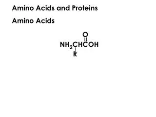
Zwitterionic form of amino acids • Under normal cellular conditions amino acids are zwitterions(dipolar ions): • Amino group = -NH3+ • Carboxyl group = -COO-
Titration Curve for Alanine • Titration curves are used to determine pKa values • pK1 = 2.4 • pK2 = 9.9 • pIAla = isoelectric point
Aliphatic R Groups • Glycine (Gly, G) - the a-carbon is not chiral since there are two H’s attached (R=H) • Four amino acids have saturatedsidechains: • Alanine (Ala, A) Valine (Val, V) • Leucine (Leu, L) Isoleucine (Ile, I) • Proline (Pro, P) 3-carbon chain connects a-C and N
Stereoisomers of Isoleucine • Ile has 2 chiral carbons, 4 possible stereoisomers
Peptide Chain Nomenclature • Amino acid“residues” compose peptide chains • Peptide chains are numbered from the N (amino) terminus to the C (carboxyl) terminus • Example: (N) Gly-Arg-Phe-Ala-Lys (C) (or GRFAK) • Formation of peptide bonds eliminates the ionizable a-carboxyl and a-amino groups of the free amino acids
Cleaving Disulfide bonds andProtecting the thiols formed • Disulfide bonds in proteins must be cleaved: • (1) To permit isolation of the PTH-cysteine during the Edman procedure (2) To separate peptide chains • Treatment with thiol compounds reduces the (R-S-S-R) cystine bond to two cysteine (R-SH) residues • Thiols are protected with iodoacetate
Further Protein Sequencing Strategies • Proteins may be too large to be sequenced completely by the Edman method • Proteases (enzymes cleaving peptide bonds) and chemicalagents are used to selectivelycleave the protein into smaller fragments • Cyanogen bromide (BrCN) cleaves polypeptides at the C-terminus of Met residues
Protease Enzymescleave specific peptide bonds • Chymotrypsin- carbonyl side of aromatic or bulky noncharged aliphatic residues (e.g. Phe, Tyr, Trp, Leu) • Trypsin - carbonyl side, basic residues (Lys,Arg). • Staphylococcus aureus V8 protease - carbonyl side of negatively charged residues (Glu, Asp). NOTE: in 50mM ammonium bicarbonate cleaves only at Glu.