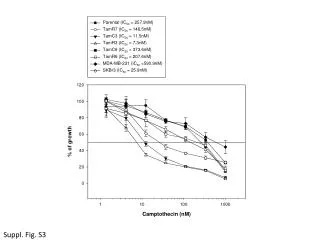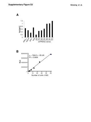
Comparative Analysis of SIRT6 in Drug-Resistant Cancer Cells and Sirt6 Knockout MEFs
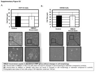
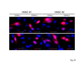
This study examines SIRT6 involvement in drug resistance by analyzing MCF-7 and MEFs with siSIRT6 or Sirt6 knockout exposed to paclitaxel and epirubicin. The experiment covers changes in FOXO3a, p53, H3K acetylation, and tubulin in cytoplasmic and nuclear cell fractions. Notable differences in protein levels observed in Sirt6 knockout MEFs are attributed to apoptosis induction. Detailed protocols for cellular lysis, fractionation, and Western blotting are included.
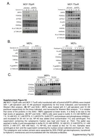

Comparative Analysis of SIRT6 in Drug-Resistant Cancer Cells and Sirt6 Knockout MEFs
E N D
Presentation Transcript
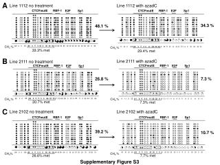
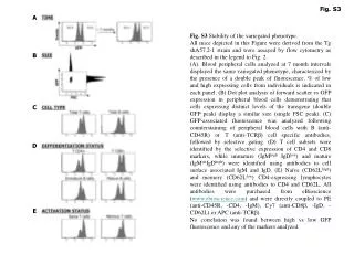
A. MCF-7EpiR MCF-7TaxR siControl siSIRT6 siControl siSIRT6 Paclitaxel (h): Epirubicin (h) 0 24 48 0 24 48 0 48 72 0 48 72 SIRT6 SIRT6 FOXO3a FOXO3a p27Kip1 p27Kip1 H3K9ac Ac-p53 H3K56ac p53 H3K9Ac Ac-α-TUBULIN H3K56Ac β-TUBULIN Ac-α-TUBULIN β-TUBULIN B. MEFs MEFs WT Sirt6-/- WT Sirt6-/- Epirubicin (h): 0 4 8 16 24 0 4 8 16 24 0 4 8 16 24 Paclitaxel (h): 0 4 8 16 24 Ac-p53 Ac-p53 p53 p53 FOXO3a FOXO3a H3K9ac H3K9ac Bim Bim P-gp(MDR1) P-gp(MDR1) β-TUBULIN β-TUBULIN C. MEFs Cytoplasmic Nuclear WT Sirt6-/- WT Sirt6-/- 0 6 24 0 6 24 0 6 24 0 6 24 FOXO3a FOXO3a (low Exp) Lamin B β-Tubulin Supplementary Figure S3. (A) MCF-7-EpiR cells and MCF-7-TaxR cells transfected with siControl/siSIRT6 siRNAs were treated with 1 μM epirubicin and 10 nMpaclitaxel respectively for the times indicated, and harvested for Western Blot analysis. (B) WT and Sirt6-/- MEFs were treated with 0.1 μM epirubicin and 10 nMpaclitaxel respectively for the times indicated, and harvested for Western Blot analysis. * It is notable that some of the proteins in the Sirt6-/- MEFs appeared at lower levels. It is likely to be due to the high levels of apoptosis. (C) WT and Sirt6-/- MEFs cells were lysed in buffer A (10 mM HEPES pH 7.4, 10 mMKCl, 0.1 mM EDTA, 0.1 mM EGTA, 2mM DTT) and protease and phosphatase inhibitors, and incubated for 20 min on ice. NP-40 was added (final concentration 1%) and centrifuged. The supernatent containing the cytoplasmic fraction was kept and frozen at -70oC. The pellet was washed in buffer A and resuspended in buffer B (10 mM HEPES, 10 mMKCl, 0.1mM EDTA, 0.1 mM EGTA, 2 mM DTT, 400 mMNaCl, 1% NP-40), and rotated at 4oC for 15 min. Samples were centrifuged at 4oC and the supernatent containing the nuclear extract collected and frozen at -70oC. The cytoplasmic and nuclear extracts were separated by SDS–PAGE gel electrophoresis, transferred to Hybond-C membranes and immunoblotted with the indicated antibodies. SupplementaryFig S3