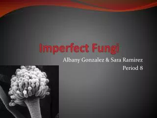
Ascomycete Anamorphs and the Imperfect Fungi
901 likes | 2.75k Vues
Ascomycete Anamorphs and the Imperfect Fungi. Mycology (Bio 594, Special Topics) M. Marshall 2013 Shippensburg University (See last slide for credits). Have mitospore types that are produced on hyphal conidiophores, or on or in structures made from aggregated hyphae = conidiomata.

Ascomycete Anamorphs and the Imperfect Fungi
E N D
Presentation Transcript
AscomyceteAnamorphs and the Imperfect Fungi Mycology (Bio 594, Special Topics) M. Marshall 2013 Shippensburg University (See last slide for credits)
Havemitospore typesthat are produced on hyphal conidiophores, or on or in structures made from aggregated hyphae = conidiomata Ascomyceteanamorphs
Most micro-fungi are first encountered as the imperfect stage Although many fungi may in fact be the imperfect asexual (anamorphic) form of a fungus with an perfect (teleomorph) stage, usually the production of the latter stage requires two opposite mating types to unite on specific substrates and/or under limited conditions. Years may elapse between the discovery of an asexual isolate and its sexual form. So many fungi that are important in human affairs are known only by their asexual designation. Some later prove to have a sexual stage and some not.
@ Deuteromycetes“deuter-” Gk., meaning “second” • > 20,000 species of fungi in 2600 genera that have no known sexual state • Most belong in phylum Ascomycota • These fungi are also called: • Anamorphic fungi • Mitosporic fungi • Conidial fungi • Imperfect fungi • Fungi imperfecti
Asexual Propagules I – Other than Conidia • Chlamydospore • 1-celled spore (usually thick-walled) designed for perennation; formed insidean existing hyphal cell • Sclerotium (pl. sclerotia) • Rounded mass of hyphae, often differentiated into rind and medulla. Usually melanized
Asexual propagules II Conidia Conidium(pl. conidia) • Non-motile spore designed for dispersal • Wide range of shape, size, color and septation among taxa (details discussed in later slides).
@ Saccardoan Spore Types & Imperfect Classification • P.A. Saccardo(1845-1920) • “SyllogeFungorum” (1882-1972)--names & descriptions of all known fungi • Developed system of classifying fungi based on type of spore (shape, septation, color)
@ Types of Deuteromycetes • Hyphomycetes—fungi that produce conidia from conidiogenous cells free on their mycelia ( on conidiophores). • May be formed on the surface of synnematalor sporodochialtypes of conidiomata • Coelomycetes—fungi that produce conidia from conidiogenous cells formed in closed or semi-closed conidiomatasuch as an ascervulus or pycnidium
Variations of Deuteromycete grouping As with all things mycological, theSaccardoansystem has been modified over the years (there are different versions) and other systems have been proposed as well:
@ SaccardoanHyphomycete (form) Families • Moniliaceae—conidiophores formed singly, hyphae and conidia pale-colored • Dematiaceae—conidiophores formed singly, hyphae and/or conidia dark-colored • Tuberculariaceae—conidiophores aggregated on cushion-like sporodochium (pl. sporodochia) • Stilbaceae—conidiophores aggregated in a synnema (pl. synnemata), an erect bundle with conidia formed at apex
Moniliales – Conidia directly on mycelium, on conidiogenous cells or conidiophores which may be separate, in clusters, or tightly packed groups. The largest and most commonly represented group. Sphaeropsidales – Conidia produced in well defined pycnidia Melanconiales – Conidia naturally produced in acervuli ; in culture possibly singly or in compact groups resembling sporodochia of the Moniliales. Mycelia Sterilia– No conidia production. Form sclerotia or other survival structures. Some authors include the conidial Oomycetes (old: Phycomycetes) here also because of their superficial similarity to the true fungi imperfects. An alternative, Saccardoan Form Orders according to Barnett*
Moniliales – Conidia directly on mycelium, on conidiogenous cells or conidiophores which may be separate, in clusters, or tightly packed groups. The largest and most commonly represented group. Moniliaceae – hyaline conidia Dematiaceae – darkly pigmented conidia (either singly or en mass). Both have conidiophores single and separate or in loose clusters. Sections: Amerosporae – conidia one-celled = amerospores Didymosporae – conidia two-celled = didymospores Phragmosporae – conidia with transverse septa only = phragmospore Dictyosporae – conidia with both transverse and oblique septation = dictyospore Scolecosporae – conidia filiform = scolecospore Staurosporae – conidia stellate or branched = staurospore Helicosporae – conidia coiled = helicospore The prefixes Hyalo- or Phaeo- are sometimes used with the above spore names to indicate hyaline or darkly pigmented respectively Barnett’s SaccardoanFamilies and Sections of the Moniliales I
Tubiculariaceae – Conidiophores compacted into a rounded or flat sporodochium which may not be produced in culture. Stilbaceae – Conidiophores compacted into synnemata, but may also produce single conidiophores of the Moniliaceous or demateaceous type (previous slide). Barnett’s*SaccardoanFamilies and Sections of the MonilialesII * H.L. Barnett was an accomplished mycologist who worked at west Virginia University. At various times he was President of the American Phytopathological Society and the Mycological Society of America. His book, Illustrated genera of Imperfect Fungi has gone through 4 editions and is still a major “Imperfect” reference today.
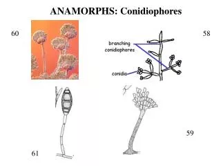
@ Conidiophores Hyphae bearing conidiogenous cells • Morphologically differentiated from vegetative hyphae (=macronematous) • Morphologically not differentiated (=micronematous)
Closed Conidiomata(Coelomycetes) AcervulusPycnidium Conidium containing structures that rupture through host epithelium. An acervulus is more open, not within walls of fungal tissue. The pycnidium is perithecium like, but contains short conidiophores and mitosporic conidia, not asci. In some ascomycete fungi the pycnidial walls develop into stroma within which true perithecia develop.
@ Open conidiomata: Synnemata • Conidiophores united at base grow in parallel to give a “tree trunk” like configuration. • Conidiogenous cells at apex. • Conidia may be produced dry or formed in a liquid matrix.
@ Open Conidiomata: Sporodochium • A compact, cushion-like aggregation of hyphae on which conidiophores are formed in a dense layer • The aggregation of hyphae is called a stroma (pl. stromata)
Saccardoan Spore Types scolecospore amerospore staurospore helicospore dictyospore phragmospore didymospore
@ More SaccardoanSpore Type Terminology • Color (prefixes) • Hyaline or bright (hyalo-) • Pigmented (phaeo-) • Shape and septation • 1-celled —amerospore • 2-celled —didymospore • Multicelled —phragmospore • Muriform —dictyospore(with both vert. & horiz. cross walls) • Filiform —scolecospore(hair-like) • Helical — helicospore • Branched — staurospore (see also slide 11)
@ Arrangement of conidia at locus • Solitary • Catenate = true chains • Seriate = false chains, spore heads • Dry spores • Wet spores (gleoid)
@ Succession of conidia • Basipetal= a chain of conidia in which new spores are formed at the base, the oldest conidia are at the apex • Acropetal = a chain of conidia with the new spores formed at the end of the chain, oldest spores are at the base. In order for this type of conidial formation to occur, the conidia must function as conidiogenous cells (e.g., Alternaria, Cladosporium)
@ Synanamorph • Two or more types of asexual spores formed by the same fungus • Example: • Ceratocystis fibriata
@ Conidiogenous Cells • A cell that forms one or more conidia • May be formed on a specialized, simple, or branched hypha called a conidiophore
@ From D. Malloch
@ Conidial Development (Ontogeny) • Blastic—blowing out of conidial initial prior to formation of delimiting septum • Thallic—conversion of segment of existing hyphae into conidia
@ Blastic versus thallic Cole, 1986
Blasticvsthallicconidiogenesis & spore separation Blastic= cross wall follows “budding”, thallic = cross wall defines the spore as separate. Schizolytic separation = septae split; rheolytic = wall of basal cell splits.
Precurrent = leaves ring-like scar on conidiogenous cell (Venturia). Phialidic = Conidia pushed out of end of conidiogenousphialid(Penicillium, Aspergilous). Retrogressive = sepatate form down the conidiophore underneath the first (?) Basauxic retrogressive-like alternative = chain of blaststic conidia where new growth is added from a mother cell below . Blastictypes: precurrent, phialidic, and retrogrssive
@ Blastic development • Holoblastic • single conidium is formed from conidiogenous locus, all wall layers involved in formation of conidium wall • Enteroblastic • more than one conidium formed from locus, only the inner wall layer(s) involved in formation of conidium wall
Enteroblasticdevelopment detail • Phialidic—a basipetal succession of conidia is formed from a fixed locus on the conidiogenous cell (phialide) collarette
Enteroblasticdevelopment detail • Annellidic—a basipetal succession of conidia formed by repeated percurrent proliferation of conidiogenous locus, leaving the distal end of locus with transverse scars (annellations)
@ More Enteroblasticdevelopment • Tretic—the inner wall of the conidiogenous cell blows out through a hole (pore) in the outer wall like a balloon to form a conidium.
Arthric = growth stops hyphae divided up by arrisingseptae. Fracture at sepatae. Alternate-arthric= some intervening cells in the arthric chain degenerate to release the others as conidia. Solitary = single large spores develop, may be multicellular. Thallic arthric, alt. arthric, and solitary
@ Arrangement of conidia at locus • Solitary • Catenate = true chains • Seriate = false chains, spore heads • Dry spores • Wet spores (gleoid)
@ Succession of conidia • Basipetal = a chain of conidia in which new spores are formed at the base, the oldest conidia are at the apex • Acropetal = a chain of conidia with the new spores formed at the end of the chain, oldest spores are at the base. In order for this type of conidial formation to occur, the conidia must function as conidiogenous cells (e.g., Alternaria, Cladosporium)
Credits This presentation has been modified from one posted on the web by Dr. Lori Carris, Washigton State University Plant Pathology Dept. from her course: Plant Path 521, Mycology.