Download
1 / 60
610 likes | 692 Vues
Explore the complexities of sensory systems, from vision and hearing to taste and smell. Learn about receptors, pathways, and how stimuli are processed. Dive into the world of somatic senses and special senses.

E N D
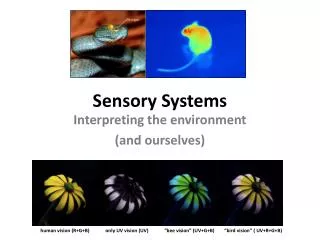
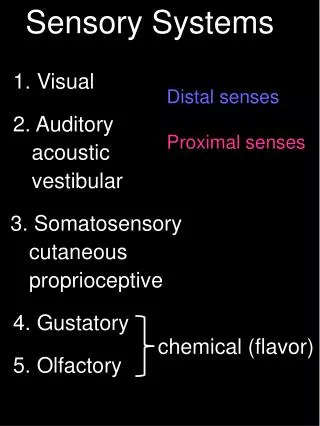
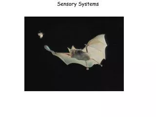
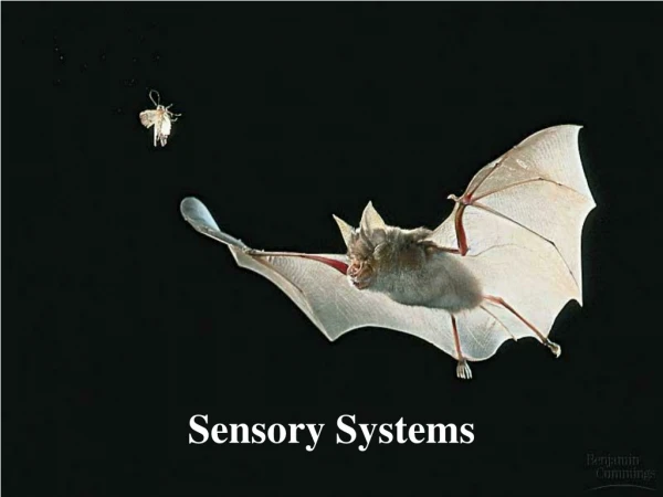
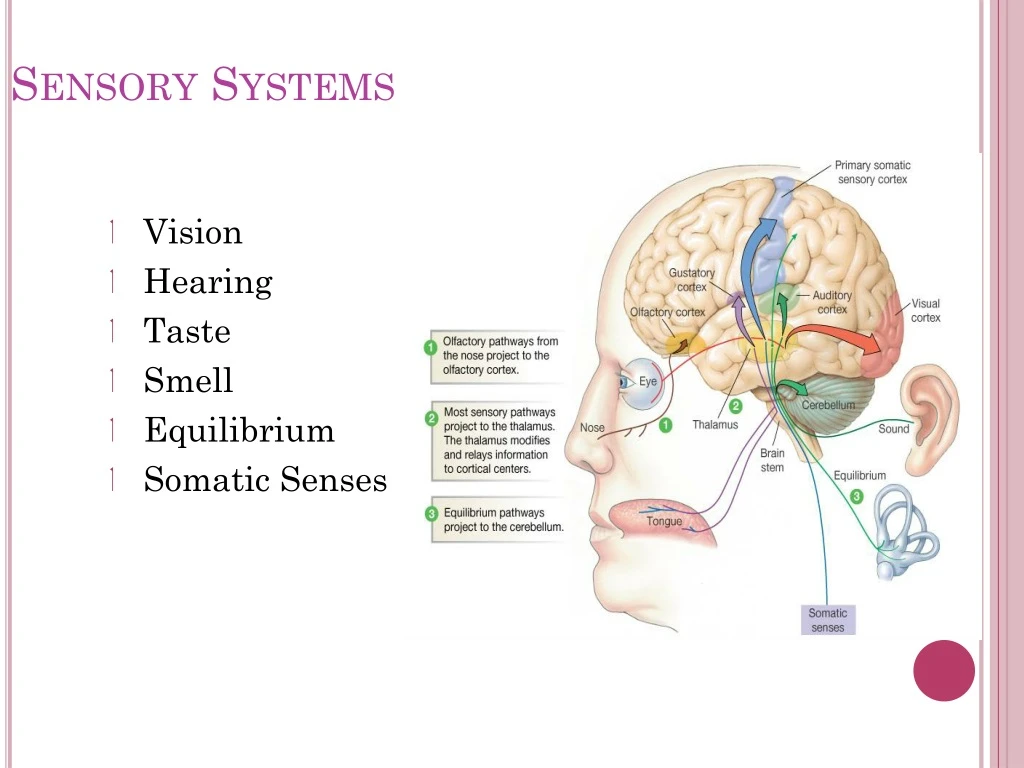
Sensory Systems • Vision • Hearing • Taste • Smell • Equilibrium • Somatic Senses
Senses • Somatic sensory • General – transmit impulses from skin, skeletal muscles, and joints • Special senses - hearing, balance, vision • Visceral sensory • Transmit impulses from visceral organs • Special senses - olfaction (smell), gustation (taste)
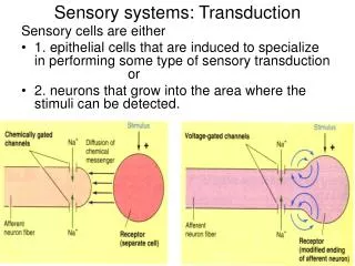
Properties of Sensory Systems • Stimulus - energy source • Internal • External • Receptors • Sense organs - structures specialized to respond to stimuli • Transducers - stimulus energy converted into action potentials • Conduction • Afferent pathway • Nerve impulses to the CNS • Translation • CNS integration and information processing • Sensation and perception – your reality
Sensory Pathways • Stimulus as physical energy sensory receptor acts as a transducer • Stimulus > threshold action potential to CNS • Integration in CNS cerebral cortex or acted on subconsciously
Classification by Function (Stimuli) • Mechanoreceptors – respond to touch, pressure, vibration, stretch, and itch • Thermoreceptors – sensitive to changes in temperature • Photoreceptors – respond to light energy (e.g., retina) • Chemoreceptors – respond to chemicals (e.g., smell, taste, changes in blood chemistry) • Nociceptors – sensitive to pain-causing stimuli • Osmoreceptors – detect changes in concentration of solutes, osmotic activity • Baroreceptors – detect changes in fluid pressure
Somatic Senses • General somatic – include touch, pain, vibration, pressure, temperature • Proprioceptive – detect stretch in tendons and muscle provide information on body position, orientation and movement of body in space
Somatic Receptors • Divided into two groups • Free or Unencapsulated nerve endings • Encapsulated nerve endings - consist of one or more neural end fibers enclosed in connective tissue
Free Nerve Endings • Abundant in epithelia and underlying connective tissue • Nociceptors - respond to pain • Thermoreceptors - respond to temperature • Two specialized types of free nerve endings • Merkel discs – lie in the epidermis, slowly adapting receptors for light touch • Hair follicle receptors – Rapidly adapting receptors that wrap around hair follicles
Encapsulated Nerve Endings • Meissner’s corpuscles • Spiraling nerve ending surrounded by Schwann cells • Occur in the dermal papillae of hairless areas of the skin • Rapidly adapting receptors for discriminative touch • Pacinian corpuscles • Single nerve ending surrounded by layers of flattened Schwann cells • Occur in the hypodermis • Sensitive to deep pressure – rapidly adapting receptors • Ruffini’s corpuscles • Located in the dermis and respond to pressure • Monitor continuous pressure on the skin – adapt slowly
Encapsulated Nerve Endings - Proprioceptors • Monitor stretch in locomotory organs • Three types of proprioceptors • Muscle spindles – monitors the changing length of a muscle, imbedded in the perimysium between muscle fascicles • Golgi tendon organs – located near the muscle-tendon junction, monitor tension within tendons • Joint kinesthetic receptors - sensory nerve endings within the joint capsules, sense pressure and position
5 Special Senses • Olfaction • Gustation • Hearing • Equilibrium • Vision
Olfactory Organs • Located in nasal cavity on either side of nasal septum Figure 17–1a
Contains • Olfactory receptors • Supporting cells • Basal (stem) cells
Olfactory Receptors • Highly modified neurons • Involves detecting dissolved chemicals as they interact with odorant-binding proteins Figure 17–1b
Taste (Gustatory) Receptors • Clustered in taste buds • Associated with epithelial projections (lingual papillae) on dorsal surface of tongue • Each taste bud contains: • basal (stem) cells • gustatory cells: • Extend taste hairs through taste pore • Survive only 10 days before replacement
Primary Taste Sensations • Sweet • Salty • Sour • Bitter • Umami • Taste vs Flavor
The Ear Figure 17–20
External Ear • Auricle • Surrounds entrance to external acoustic canal • Protects opening of canal • Provides directional sensitivity • External acoustic canal • Canal that runs from auricle to tympanic membrane • Tympanic membrane (Eardrum) • Is a thin, semitransparent sheet • Separates external ear from middle ear
External Ear Figure 17–20
Ceruminous Glands • Integumentary glands along external acoustic canal • Secrete waxy material (cerumen): • keeps foreign objects out of tympanic membrane • slows growth of microorganisms in external acoustic canal
3 Auditory Ossicles • Malleus (hammer) • Incus (anvil) • Stapes (stirrup)
Vibration of Tympanic Membrane • Converts arriving sound waves into mechanical movements • Auditory ossicles conduct vibrations to inner ear
Inner Ear Figure 17–20
Inner Ear • Contains fluid • Subdivided into: • vestibule • semicircular canals • cochlea
Parts to Inner Ear • Vestibular Complex • Combination of vestibule and semicircular canals • Vestibule • Receptors provide sensations of gravity and linear acceleration • Semicircular Canals • Contain semicircular ducts • Receptors stimulated by rotation of head • Cochlea • Contains cochlear duct • Receptors provide sense of hearing
Equilibrium • Sensations provided by receptors of vestibular complex • https://www.youtube.com/watch?v=KuiNueVxdec
Sound • Consists of waves of pressure through air or water
Wavelength • Distance between 2 adjacent wave troughs • Frequency • Number of waves that pass fixed reference point at given time • Physicists use term cycles instead of waves • Hertz (Hz) • Number of cycles per second (cps)
Pitch • Our sensory response to frequency • Increased frequency results in a higher pitch • Decreased frequency results in a lower pitch
Amplitude • Intensity of sound wave • Sound energy is reported in decibels
The Power of Sounds Table 17–1
How do we hear? • Sound waves enter external acoustic canal • Soundwaves vibrate the tympanic membrane • Vibrations are transferred to and through the auditory ossicles • Vibrations are transferred to fluid in cochlea • Nerve endings pick up vibrations and send signal to brain
Accessory Structures of the Eye • Eyelids • Superficial epithelium of eye • Structures associated with production, secretion, and removal of tears
Eyelids (Palpebrae) • Continuation of skin • Blinking keeps surface of eye lubricated, free of dust, and debris
Eyelashes • Robust hairs that prevent foreign matter from reaching surface of eye
Tarsal Glands • Associated with eyelashes • Secrete lipid–rich product that helps keep eyelids from sticking together • Contribute to gritty deposits that appear after good night’s sleep
Conjunctiva • Epithelium covering inner surfaces of eyelids and outer surface of eye • Conjunctivitis • Results from damage to conjunctival surface Figure 17–3b
Cornea • Transparent part of outer fibrous layer of eye
Lacrimal Gland(Tear Gland) • Secretions contain lysozyme, an antibacterial enzyme • Lubricates, cleanses, disinfects eye
Orbital Fat • Cushions and insulates eye Figure 17–4c
Eyeball • Is hollow • Is divided into 2 cavities: • large posterior cavity • smaller anterior cavity
Outer Surface of Eye • Sclera (white of eye) • Cornea
Middle Layer of Eye • Includes: • iris • ciliary body • Iris • Contains muscle fibers • Changes diameter of pupil • Ciliary body • Assist in changing shape of lens for focusing images
Lens • Lies posterior to cornea • Forms anterior boundary of posterior cavity
Inner Layer of Eye (Retina) • Outer pigmented part • Inner neural part: • contains visual receptors and associated neurons