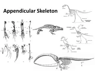
Appendicular Skeleton
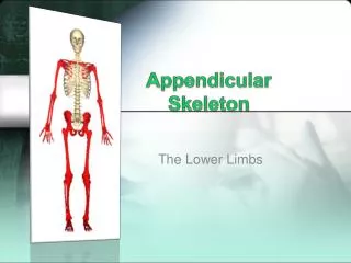
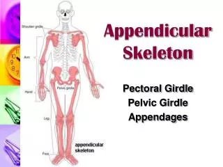
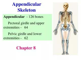
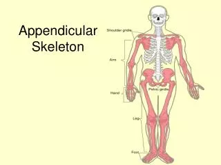
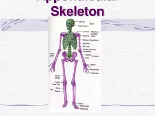
Appendicular Skeleton. The appendicular skeleton is made up of the appendages - the legs and arms. The superior appendicular skeleton is the pectoral (shoulder) girdles - it attach the upper limbs to the body trunk.

Appendicular Skeleton
E N D
Presentation Transcript
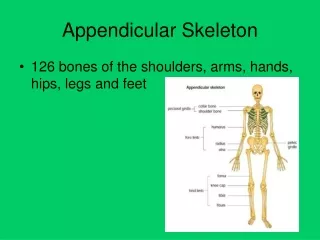
Appendicular Skeleton • The appendicular skeleton is made up of the appendages - the legs and arms. • The superior appendicular skeleton is the pectoral (shoulder) girdles - it attach the upper limbs to the body trunk. • The inferior appendicular skeleton is the pelvic girdle -it secures the lower limbs.
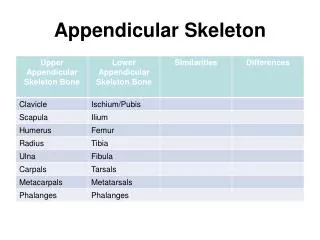
Pectoral Girdles (Shoulder Girdles) • The pectoral girdles consist of the anterior clavicles and the posterior scapulae • They attach the upper limbs to the axial skeleton in a manner that allows for maximum movement • They provide attachment points for muscles that move the upper limbs
Clavicles (Collarbones) • The clavicles (2) are slender, curved long bones lying across the superior thorax • The acromial (lateral) end articulates with the scapula, and the sternal (medial) end articulates with the sternum • They provide attachment points for numerous muscles, and act as braces to hold the scapulae and arms out laterally away from the body
Scapula (Shoulder Blades) • The scapulae (2) are triangular, flat bones lying on the dorsal surface of the rib cage, between the second and seventh ribs • Scapulae have three borders - superior, lateral, medial • Major markings include the spine, the acromion, the glenoid cavity and the coracoid process
The Upper Limb • The upper limb consists of the arm (brachium), forearm (antebrachium), and hand (manus) • Thirty-seven bones form the skeletal framework of each upper limb
The Humerus (Arm) • The humerus is the sole bone of the arm • It articulates with the scapula at the shoulder, and the radius and ulna at the elbow • Major markings • Proximal humerus includes the head, anatomical and surgical necks, greater and lesser tubercles, and the intertubercular groove • Distal humerus includes the capitulum, trochlea, medial and lateral epicondyles.
The Radius and Ulna (Forearm) • The bones of the forearm are the radius and ulna • They articulate proximally with the humerus and distally with the wrist bones • They also articulate with each other proximally and distally at small radioulnar joints • Interosseous membrane connects the two bones along their entire length
The Radius • The radius lies opposite (lateral to) the ulna and is thin at its proximal end, widened distally • In anatomical position it is the bone closest to the thumb • The superior surface of the head articulates with the capitulum of the humerus • Medially, the head articulates with the radial notch of the ulna • Major markings include the radial tuberosity, ulnar notch, and styloid process • During wrist rotation, the distal end crosses the ulna
The Ulna • The ulna lies medially in the forearm and is slightly longer than the radius • Forms the major portion of the elbow joint with the humerus • Its major markings include the olecranon, coronoid process, trochlear notch, radial notch, and the styloid process
The Hand • Skeleton of the hand contains wrist bones (carpals), bones of the palm (metacarpals), and bones of the fingers (phalanges)
Pelvic Girdle (Hip) • The hip is formed by a pair of hip bones (os coxae, or coxal) • Together with the sacrum and the coccyx, these bones form the bony pelvis • The pelvis • Attaches the lower limbs to the axial skeleton with the strongest ligaments of the body • Transmits weight of the upper body to the lower limbs • Supports the visceral organs of the pelvis
Comparison of Female and Male Pelvic Structure • Female pelvis • Tilted forward, adapted for childbearing • True pelvis defines birth canal • Cavity of the true pelvis is broad, shallow, and has greater capacity • Male pelvis • Tilted less forward • Adapted for support of heavier male build and stronger muscles • Cavity of true pelvis is narrow and deep
Carpus - Wrist • Consists of eight bones connected by ligaments - 2 rows of 4 • Scaphoid, lunate, triquetral, and pisiform proximally • Trapezium, trapezoid, capitate, and hamate distally
Metacarpus - Palm • Five numbered (1-5) metacarpal bones radiate from the wrist to form the palm - the heads form the knuckles • Their bases articulate with the carpals proximally, and with each other medially and laterally • Heads articulate with the phalanges
Phalanges (Fingers) • Each hand contains 14 miniature long bones called phalanges • Fingers are numbered 1-5, beginning with the thumb (pollex) • Each finger (except the thumb) has three phalanges – distal, middle, and proximal - the thumb has no middle phalanx
Ilium • The ilium is a large flaring bone that forms the superior region of the coxal bone • It consists of a body and a superior winglike portion called the ala • The broad posterolateral surface is called the gluteal surface • The auricular surface articulates with the sacrum (sacroiliac joint) • Major markings include the iliac crests, four spines, greater sciatic notch, iliac fossa, arcuate line, and the pelvic brim
Ischium • The ischium forms the posteroinferior part of the hip bone • The thick body articulates with the ilium, and the thinner ramus articulates with the pubis • Major markings include the ischial spine, lesser sciatic notch, and the ischial tuberosity
Pubis • The pubic bone forms the anterior portion of the hip bone • It articulates with the ischium and the ilium • Major markings include superior and inferior rami, the pubic crest, pubic tubercle, pubic arch, pubic symphysis, and obturator foramen (along with ilium and ischium)