Download
1 / 17
170 likes | 299 Vues
This comprehensive overview of knee injuries for athletic trainers covers essential anatomy, including bones, muscles, cartilage, ligaments, and bursa. It discusses the functional anatomy of the knee, key motions, and evaluation techniques such as history-taking, observation, and special tests. Detailed sections on MCL, LCL, ACL, and PCL sprains present their mechanisms, signs, symptoms, and treatment strategies like RICER and rehabilitation. Additional topics include meniscal lesions, patellar dislocation, patellofemoral syndrome, and apophyseal injuries, emphasizing the importance of accurate assessment and targeted interventions.

E N D
Athletic InjuriesATC 222 The Knee Chapter 16
Anatomy • bony • muscular • cartilage • ligaments • bursa • etc
Functional Anatomy • Motions • flexion/extension • rotation
Evaluation • History • Observation • Palpation • Special Tests
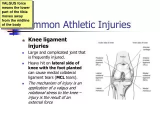
MCL Sprain • Mechanism • lateral force • external tibial rotation • hyperextension • More severe and common than LCL • part of capsule • attaches to medial meniscus • Signs and Symptoms • pain over MCL • none to moderate edema/effusion • possible instability • possible ROM and strength loss • pain with passive extension and external tibial rotation
MCL Sprain Treatment • RICER • Crutches? • Immobilization? • Meniscal involvement? • Rehabilitation
LCL Sprain • Mechanism • varus force, internal tibial rotation • Less prevalent that MCL • no meniscal attachment • action of popliteus muscle • common peroneal nerve damage? • Signs and Symptoms • pain over LCL • none to mild edema/effusion • possible instability • possible ROM and strength loss • pain with passive extension and internal tibial rotation • Treatment
ACL Sprain • Mechanism • external tibial rotation with valgus • internal tibial rotation • hyperextension • deceleration • Most common knee ligament to be seriously injured • Signs and Symptoms • heard/felt a “pop” • rapid effusion/hemarthrosis (1-2 hours) • knee “gives out” • instability
ACL Sprain Treatment • RICER • Crutches? • Immobilization? • Conservative vs surgical intervention • commonly associated with meniscal tear
PCL Sprain • Mechanism • force to anterior tibia with knee flexed • hyperflexion • rotation • Signs and Symptoms • “pop” • effusion • instability • Treatment • usually non-operative
Meniscal Lesions • Mechanism • rotation while weight bearing • Acute MCL or ACL sprain • chronic knee instability or degeneration • Signs and Symptoms • clicking, catching, locking • slow developing effusion • pain on joint line • chronic effusion • Treatment • healing rate? • Arthroscopic surgery • removal vs repair
Patellar Subluxation/Dislocation • Mechanism • external tibial rotation • valgus • functional/structural deviations • Signs and Symptoms • deformity • positive apprehension sign • effusion • pain on medial patellar border • Treatment • Reduction • immobilization and crutches? • RICER • rehabilitation
Patellofemoral Syndrome • Structural/functional deviations • Signs and Symptoms • medial peripatellar pain • pain with stairs • crepitus • pain with stairs/prolonged sitting • Treatment • symptomatic • correct functional/structural deviations
Apophyseal Injuries(apophysitis) • Apophysis = Traction Epiphysis • Types • Osgood-Schlatter’s Syndrome • Sinding-Larson-Johanson Syndrome • Signs and Symptoms • pain at tendon attachment • tibial tubercle enlargement • pain with prolonged sitting or pressure • Treatment • symptomatic • flexibility • activity modification • straps and sleeves
Other Injuries • Tendonitis • Osteochondral defects • ITB syndrome
Special Tests of the Knee • tibial/fibular compression/percussion • patellar apprehension • ballotable patella and stroke test • patellar excursion and compression
Other Special Considerations • leg length discrepancy • Mechanics of lower extremity • Q-Angle • 10 males, 15 females • over 20 is abnormal