Characterization of Amyloid Oligomers and Fibrils using TEM Analysis
Detailed methods for TEM analysis of amyloid structures, including sample preparation, staining, and data interpretation, with illustrative figures.

Characterization of Amyloid Oligomers and Fibrils using TEM Analysis
E N D
Presentation Transcript
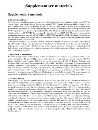
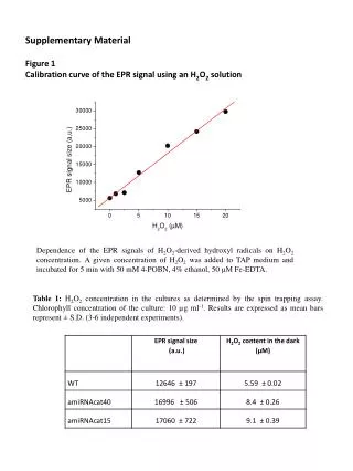
Supplementary Methods TEM (transmission electron microscopy). TEM analysis were performed on EF-TEM LEO 912AB (Carl Zeiss Inc., Germany, Korea Basic Science Institute, Chuncheon) operated at 120 kV. Amyloid oligomers and fibrils were negatively stained using uranyl acetate. The samples were absorbed on formvar-carbon coated grid (300 mesh, Electron Microscopy Science) for 1 min and negatively stained for 1 min with 1% uranyl acetate solution. After washing with distilled water, the sample-coated grid was dried in air and then processed for TEM analysis.
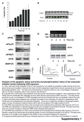
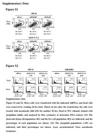
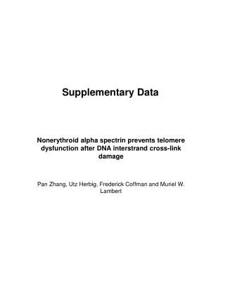
Figure Legend for Supplementary Data Supplementary Figure S1. Purification of rhGCPII protein and inhibition of GCPII activity by inhibitors. A, Coommassie-stained gel and western blot of rhGCPII preparations are shown.The lysate of SF21 cells infected with GCPII showed multiple bands including a ~100 kDa band. After purification by Ni-NTA column and dialysis we could detect oneband which was confirmed as GCPII by western blotting with anti-human GCPII antibody (arrow). B, NAAG cleavage activity of purified rhGCPII was measured. 10 μM [3H]-labeled NAAG was incubated with rhGCPII (15 ng/ml). The released [3H]-labeled glutamate was isolated by ion exchange chromatography and quantified. Pre-incubation of the enzyme with 1 mM 2-PMPA, 2 mM Phen, and 20 mM EDTA inhibited cleavage of NAAG. Supplementary Figure S2. MALDI-TOF MS analysis of Aβ peptide cleavage by rhGCPII. Aβ1-40 (20 μM) was incubated at 37°C either with increasing amounts of rhGCPII for 24 h (A) or with rhGCPII (15 ng/ml) for the time indicated (B). Aβ1-42 (20 μM) was also cleaved by rhGCPII but was not cleaved by heat-treated rhGCPII. Supplementary Figure S3. Preparation of small-, medium- and large-sized Aβ oligomers. A, Aβ oligomers were prepared from the Aβ1-42 (80 μM) solution by incubating for 24 h at 4C, room temperature or 37C, while Aβ fibrils by incubating the Aβ1-42 (40 μM) solution for 6 days at 37°C. Samples were centrifuged and analyzed by western blotting using with anti-Aβ antibody (6E10). B, Transmission electron microscopy images of the Aβ42 oligomer and fibril preparations are shown. (bar = 100 nm) Supplementary Figure S4. Comparison of neurotoxicity induced by different forms of Aβs. Primary rat cortical neuronal cultures were treated with Aβ1-14 (10 μM) or Aβ1-42 monomers (10 μM) for 60 h and the viability of cells was examined by MTT assay. Supplementary Figure S5. Measurement of rhGCPII stability. In order to examine the stability of GCPII rhGCPII (15 ng/ml) was incubated at 37C for 1, 3, or 6 days and assayed for NAAG cleavage activity (A) or Aβ1-40 cleavage activity by MALDI-TOF analysis (B). Incubation of rhGCPII for 1- 3 days caused no significant reduction while that for 6 days resulted in ~ 20% reduction. In contrast, all three rhGCPII preparations showed no reduction in Aβ cleavage activity. * p< 0.05
Supplementary Figure 5. B A * 1-40 1-40 1-40