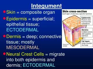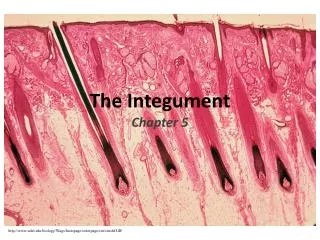
Integumentary System Layers and Structures
370 likes | 453 Vues
Dive into the intricate layers and structures of the integumentary system, including the epidermis, dermis, and integumentary accessory structures like hair follicles, sweat glands, and sebaceous glands. Learn about the functions and features of each component in detail.

Integumentary System Layers and Structures
E N D
Presentation Transcript
Organization of the Epidermis: Figure 5–2
Layers of the Epidermis Top: Free surface of skin - stratum corneum - stratum lucidum - stratum granulosum - stratum spinosum • stratum germinativum Bottom: Basal lamina
A note on thick vs. thin skin • Thick skin has an extra layer (lucidum) but that is NOT the reason that it is thicker than thin skin. • Real reason is the other layers are thicker in thick skin than in thin skin.
The Dermis • Deeper part of cutaneous layer • Located between epidermis and subcutaneous layer • Anchors epidermal accessory structures (hair follicles, sweat glands) • Has 2 components: • outer papillary layer • deep reticular layer
The Papillary Layer • Consists of areolar tissue • Contains smaller capillaries, lymphatic vessels, and sensory neurons • Has dermal papillae projecting between epidermal ridges
The Reticular Layer • Consists of dense irregular connective tissue • Contains larger blood vessels, lymph vessels, and nerve fibers • Contains collagen and elastic fibers
Integumentary Accessory Structures • Hair, hair follicles, sebaceous (oil) glands, sweat glands, and nails: • are derived from embryonic epidermis • are located in dermis • project through the skin surface
The Hair Follicle • Is located deep in dermis • Is made of epidermal tissue (with connective tissue around the outside) • Produces nonliving hairs • Is wrapped in a dense connective-tissue sheath • Base is surrounded by sensory nerves
Structures of Hair and Follicles Figure 5–9a
Accessory Structures of Hair • Arrector pili: • involuntary smooth muscle • causes hairs to stand up • produces “goose bumps” • Sebaceous glands: • lubricate the hair • control bacteria
Inside the Follicle Figure 5–9b
Exocrine Glands in the skin • Sebaceous glands and follicles (oil glands): • holocrine glands • secrete sebum • Sweat glands: • merocrine glands • watery secretions
Types of Sebaceous Glands • Sebaceous glands: • associated with most hair follicles (on head and body) • Sebaceous follicles: • discharge directly onto skin surface • found on face and trunk • when clogged acne
Types of Sweat Glands • Apocrine: • found in armpits, around nipples, and groin • Merocrine: • more numerous, widely distributed on body surface • especially on palms and soles (thick skin) Both are actually merocrine
“Apocrine” Sweat Glands • Merocrine secretions, not apocrine • Associated with hair follicles in groin, nipples, and axillae (armpits) • Become active at puberty • Produce sticky, cloudy secretions (thick sweat) that breaks down and causes odor
Merocrine Sweat Glands • Also called eccrine glands: • coiled, tubular glands • discharge directly onto skin surface • sensible perspiration for cooling (thin sweat) • water, salts, and organic compounds
Sweat Glands of the Skin Merocrine Apocrine
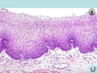
Epidermis What to look for: • Usually darkest between stratum germinativum and stratum granulosm (granulosm often a dark meandering line) • Keratinized cells (s. corneum) often lift off the underlying layers • S. germinativum along basal lamina, along with melanocytes
What to look for • Papillary layer • has ridges • is areolar • Just under basal lamina • Reticular layer • much thicker • Dense irregular CT • Hypodermis • Loose CTP
What to look for Found in most skin Coiled, tubular Small lumens in cross section Have duct that goes all the way to the epidermal surface and ends in sweat pore Smaller than apocrine, don’t extend as deep into dermis Merocrine sweat gland
Apocrine sweat gland What to look for: • Associated with hair follicle • Only in nipples, groin, armpit • Large lumens • Deeper in dermis than merocrine
Hair What to look for: • Follicles are rarely complete • Can often see root, papilla at base of hair • Arrector pilli muscle at an angle • Associated glands (which are?)
Sebaceous glands What to look for: • Associated with hair follicle • Found most everywhere hair follicles are found in skin • Look like cauliflower (maybe?)
Sebaceous follicle What to look for: • Also look like cauliflower • Found on face and trunk only • NOT associated with hair follicle • Have duct that opens onto skin surface