Download
1 / 110
1.1k likes | 1.14k Vues
Explore the structure and classification of bones, including long, short, flat, irregular, and sesamoid bones. Learn about the microscopic anatomy of bones and the processes of bone development and growth. Discover the role of nutrition and hormones in bone health.

E N D
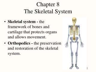
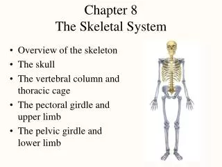
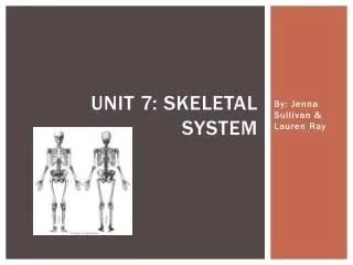
Chapter 7 - The Skeletal System Individual bones are the organs of the skeletal system. A bone contains very active tissues.
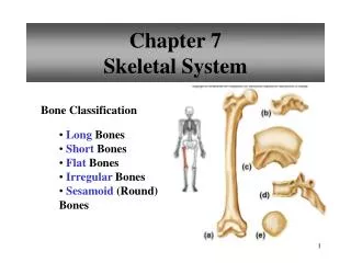
Bone classification • Classified according to their shapes: • Long - longitudinal axis and expanded ends Ex: forearm and thigh bones • Short - cube like with lengths and widths about equal Ex: wrist and ankle
Flat - platelike with broad surfaces Ex: ribs, scapula, some skull bones • Irregular - variety of shapes, usually connected to several other bones. Ex: vertebrae and facial bones • Sesamoid - round, usually small and nodular embedded within tendons next to joints where tendons are compressed Ex: kneecap
Parts of a long bone • Epiphysis at each end that articulates (forms a joint) with another bone • Outer surface of epiphysis covered with a layer of hyaline cartilage called articular cartilage • The shaft of a bone is called the diaphysis • Except for the articular cartilage, a bone is covered by fibrous tissue called the periosteum.
Wall of diaphysis made of compact bone that has a continuous matrix with no gaps. Epiphysis made of spongy bone that has irregular interconnecting spaces between bony plates (trabeculae). Both compact and spongy bone are strong and resist bending. The diaphysis contains a medullary cavity filled with marrow.
Microscopic structure Osteocytes (bone cells) located in lacunae which form concentric circles around osteonic canals
Compact bone Osteocytes and layers of intercellular material concentrically clustered around an osteonic canal to form osteons cemented together. Osteonic canals contain blood vessels that nourish the cells of osteons. Perforating canals connect osteonic canals transversely and communicate with the bone's surface and the medullary cavity.
Spongy bone Composed of osteocytes and intercellular material but the cells don not aggregate around osteonic canals Cells lie within trabeculae Diffusion from the surface of thin bony plates nourishes cells of spongy bones.
Bone Development and Growth Bones form by replacing existing connective tissue in an embryo and continue to grow and develop into adulthood.
Intramembraneous Ossification Flat, broad bones in skull Membranelike layers of unspecialized connective tissue appear at future bone sites Unspecialized connective tissue cells arrange blood vessels in layers Connective tissue cells differentiate into osteoblasts, which deposit spongy bone
Osteoblasts become osteocytes when a bony matrix completely surrounds them. Connective tissue on the surface of each developing structure forms a periosteum. Osteoblasts on the inside of periosteum deposit compact bone over the spongy bone.
Process of bone formation is called ossification. Ossification timeline - see table 7.2 page 209.
Endochondral Ossification • Masses of hyaline cartilage form models of future bone • Cartilage tissue breaks down, periosteum develops • Blood vessels and osteoblasts invade tissue • Osteoblasts form spongy bone • Osteoblasts deposit a thin layer of compact bone
Osteoblasts become osteocytes when bony matrix surrounds them. • Osteocytes are mature bone cells
Growth of an endochondral bone Primary ossification center appears in the diaphysis and bone develops towards the ends Secondary ossification centers appear in the epiphysis and spongy bone forms around them An epiphyseal disk (band of cartilage) remains between the primary and secondary ossification centers.
An epiphyseal disk consists of 4 layers of cells • resting cells - anchor disk to bony tissue of epiphysis • young reproducing cells - undergo mitosis • older enlarging cells - enlarge and thicken the disk causing bone to lengthen • dying cells - thin layer
Long bones continue to lengthen until the epiphyseal disks are ossified. Growth in thickness occurs as compact bone is deposited beneath the periosteum. Osteoclasts erode bone tissue inside which later fills with marrow. Throughout life osteoclastsresorb bone tissue (resorption) and osteoblasts replace bone (deposition). 3% -5% of calcium exchanged each year. The total mass of bone remains nearly constant.
Nutrition • Vitamin D necessary for proper calcium absorption • Bones deformed without - rickets in children and osteomalacia in adults • Vitamin A needed for bone resorption Lack of retards bone development • Vitamin C needed for collagen synthesis Lack of results in bones abnormally slender and fragile
Sunlight • Converts dehydrocholesterol produced by cells in the digestive tract into vitamin D. • Vitamin D allows calcium to be absorbed
Hormones Insufficient secretion of pituitary growth hormone may result in dwarfism. excessive secretion of GH may result in giantism (>8ft.) or acromegaly( hands, feet, and jaw enlarge). Deficiency of thyroid hormone delays bone growth. Male and female sex hormones promote bone formation and stimulate ossification of the epiphyseal disks.
Physical stress Skeletal muscle contractions pulls on bone attachments which stimulates bone tissue to thicken and strengthen.
Support and Protection • Body movevement • Blood Cell Formation (hematopoiesis) Begins in the yolk sac, outside the embryo Occurs later in the liver, spleen and bone marrow
Red marrow houses developing red blood cells, white blood cells, and blood platelets. Red in color due to hemoglobin. In adults found in spongy bone of the skull, ribs, sternum, clavicles, vertebrae, and pelvis. Yellow marrow stores fat and replaces red marrow as we age.
Inorganic salt storage • Bone matrix is made of collagen and inorganic mineral salts. • Salts are about 70% of matrix by weight. • Most salt is calcium phosphate in the form of hydroxyapatite. • Calcium is needed for blood clot formation, nerve impulse conduction, and muscle cell contraction
When blood is low in calcium osteoclasts resorb bone, releasing calcium salts. When blood is high in calcium osteoblasts are stimulated to form bone tissue and store calcium salts. Bone also stores small amounts of sodium, magnesium, potassium, and carbonate ions.
Number of bones • Usually a human has 206 bones, but the number may vary. • Extra bones in sutures (flat bones in skull grow together) are called sutural bones.
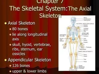
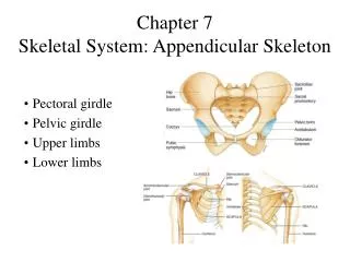
Divisions of the skeleton Axial- skull, hyoid bone (supports tongue), vertebral column, and thorax Appendicular - pectoral girdle, upper limbs, pelvic girdle, and lower limbs.
Skull Consists of twenty-two bones, which include eight cranial bones, thirteen facial bones, and one mandible.
Cranium • Encloses and protects the brain, and provides attachments for muscles. • Some bones contain air-filled sinuses that help reduce the weight of the skull. • Cranial bones include the frontal bone, parietal bones, occipital bone, temporal bones, sphenoid bone, and ethmoid bone.
Facial skeleton Form the basic shape of the face and provide attachments for muscles. Consist of 13 immovable bones and a movable lower jawbone (mandible). Facial bones include the maxillary bones(2), palatine bones(2), zygomatic bones(2), lacrimal bones(2), nasal bones(2), vomer bone, inferior nasal conchae(2) and mandible.
Infantile skull Incompletely developed bones, connected by fontanels (soft spots) enable the infantile skull to change shape slightly during childbirth. Small face with a prominent forehead and large orbits. Bones are thin and somewhat flexible and less easily fractured than adults.
Vertebral Column • Extends from the skull to the pelvis and protects the spinal cord. • Composed of vertebrae separated by intervertebral disks. • Vertebrae connected to one another by ligaments. • An infant has thirty-three vertebral bones - 5 eventually fuse to become the sacrum and 4 fuse to become the coccyx. • An adult has twenty-six vertebrae. • The vertebral column has four curvatures -cervical, thoracic, lumbar, and pelvic.