Download
1 / 1
10 likes | 128 Vues
This study explores the role of Wnt/β-Catenin signaling in enhancing radiation resistance of mammary stem cells. Results indicate radiation increases stem-like progenitor cells in mammary cell lines, highlighting possible targets for molecular therapeutics.

E N D
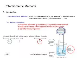
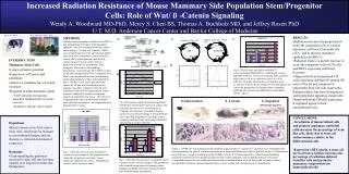
pMSCV- IRES - GFP pMSCV- -cateninmut - IRES - GFP 5’ LTR 5’ LTR Amp + Amp + GFP -cateninmut IRES-GFP IRES-GFP 3’ LTR 3’ LTR Increased Radiation Resistance of Mouse Mammary Side Population Stem/Progenitor Cells: Role of Wnt/ ß -Catenin Signaling Wendy A. Woodward MD-PhD, Mercy S. Chen BS, Thomas A. Buchholz MD, and Jeffrey Rosen PhD U.T. M.D. Anderson Cancer Center and Baylor College of Medicine Baylor cow parade M. D. Anderson Cancer Center • RESULTS: • Radiation increases the proportion of stem-like progenitor cells in a mouse mammary cell lines, CommaD cells (CD), and in primary mammary epithelial cells(MECs) • Radiation leads to a greater increase in stem-like progenitor cells in CD cells and MECs expressing stabilized • ß-catenin • Outgrowths from transplanted CD cells expressing stabilized ß-catenin fill more of the fat pad compared to outgrowths from CD cells expressing • ß-galactosidase, but form disorganized and hyperplastic appearing outgrowths. Outgrowths from CD cells expressing • ß-engrailed appear to lack myoepithelial cells • METHODS • Primary mouse mammary epithelial cells (MECs) and immortalized Comma D (CD) mammary epithelial cells were irradiated in 60 mm culture dishes using a 137cesium cell irradiator. MECs were isolated from 6-8 week old wild type Balb/c mice, from mice containing a floxed exon III ß-catenin allele, which generates stabilized ß-catenin upon excision in culture with an adenovirus-driven Cre recombinase, from transgenic Wnt-1mice with mammary hyperplasias, from wild type (WT) MECs in mice of the same background as Wnt-1 transgenic mice. MECs were maintained in stem cell promoting media after plating for 2 days and irradiated on day 4. On day 5 cells were trypsinized and stained for 60 min with Hoechst dye prior to analysis using flow cytometry. CD cells were transduced with control ß-galactosidase, amino-terminal, stabilized ß-catenin, or dominant-negative ß-engrailed retroviruses. CD cells were transplanted into the cleared fat pads of Balb/c mice 48h after transduction, and outgrowths were biopsied after 8 weeks. A. A. C. • INTRODUCTION • Mammary Stem Cells: • Long replicative potential • Capacity to self-renew and proliferate • Attractive candidate for cell origin of cancer • Required in adult mammary gland • Fulfill demands of pregnancy-dependent epithelial turnover (tissue renewal) • Respond to damage (tissue repair) • Hypothesis: • Breast cancers arise from cancer stem cells, which may be resistant to conventional therapy, and are therefore a critical determinant of recurrence • Rationale: • Understanding the pathways involved in stem cell survival may identify new targets for molecular therapeutics B. Harada et al., EMBO J . 18:5931, 1999 Fig 4. CD cells were transduced with MSCV vector containing IRES-GFP or a stabilized ß -catenin and IRES-GFP(A). Cells were sorted by flow cytometry and positive cells were cultured to enrich for GFP+ cells (B). Clinically relevant doses of radiation lead to a greater increase the percentage of side population cells (stem-like progenitors) in primary mouse mammary epithelial cells expressing stabilized ß-catenin than in control cells (C). B. ß-Galactosidase ß -Catenin ß -Engrailed (Dominant negative) Fig. 2 Floxed exon III ß-catenin allele generates stabilized ß-catenin upon excision in culture with an adenovirus-driven Cre recombinase (A). Clinically relevant doses of radiation lead to a greater increase the percentage of side population cells (stem-like progenitors) in primary mouse mammary epithelial cells expressing stabilized ß-catenin than in control cells (B). • CONCLUSIONS: • Irradiation of immortalized cells and primary mammary epithelial cells increases the percentage of stem-like cells, likely due to stem cell radioresistance relative to the differentiated cells • Expression of ß-Catenin, a stem cell survival factor, further increases the percentage of radiation-induced stem-like cells and promotes mammary outgrowth from immortalized cells Figure 5. 10,000 CD cells transduced with stabilized ß-galactoidase ß -catenin or ß -engrailed were transplanted into cleared mammary fat pads. ß -catenin outgrowths were larger and filled more of the fat pad at 8 weeks. ß -Galactosidase outgrowths were roughly normal appearing on H&E sections. ß -Catenin outgrowths contain normal branching and ductal elements but were highly disorganized and contain abnormal areas of undifferentiated cells (inset). ß -engrailed outgrowths were small and characterized by marked dilation of of the ducts with irregular or absent myoepithelial cells(inset: immunohistochemistry of same section with smooth muscle actin antibody). Fig. 1 Clinically relevant doses of radiation increase the percentage of side population cells (stem-like progenitors) in primary mouse mammary epithelial cell culture. Magnitude of increase varies by mouse strain. Fig. 3 Clinically relevant doses of radiation lead to a greater increase the percentage of side population cells (stem-like progenitors) in primary mouse mammary epithelial cells from transgenic Wnt-1 mice with mammary hyperplasias.


![[virtual] cells](https://cdn1.slideserve.com/3553683/slide1-dt.jpg)