Distinct Biotinylated Membrane Protein Profile in Cultured Hippocampal Neurons
Surface proteins of hippocampal neurons were biotinylated and analyzed, revealing differences in protein profiles. This experiment helps understand neuronal surface protein dynamics.

Distinct Biotinylated Membrane Protein Profile in Cultured Hippocampal Neurons
E N D
Presentation Transcript
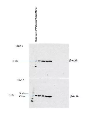
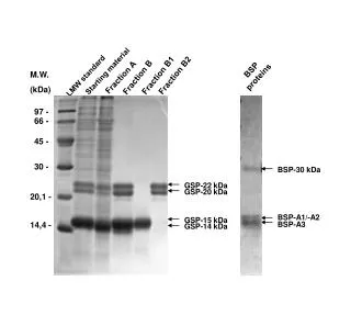
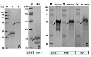
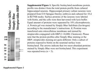
Supplemental Figure 1. Specific biotinylated membrane protein profile was distinct from the total protein profile from cultured hippocampal neurons. Hippocampal primary culture neurons were prepared from E18 Sprague Dawley embryos and cultured for 10 d in B27NB media. Surface proteins of the neurons were labeled with biotin, and the cells were then harvested with lysis buffer. Equal amount of proteins were separated by 10% electrophoresis. A, Protein gel was stained by Simply Blue SafeStain (Invitrogen) according to the manufacturer’s instructions. B, Proteins were transferred onto nitrocellulose membrane and stained by streptavidin conjugated with HRP (1:10,000; Chemicon). Please note that the protein profiles after Simply Blue staining or the HRP--streptavidin staining are quite different. Only a subset of proteins exposed to biotin on the neuronal surface were biotinylated. The arrows indicate that two most abundant proteins stained by Simply Blue were not biotinylated. This experiment was repeated twice (n = 3). Streptavidin-HRP Marker (kDa) Simply Blue 250 148 98 64 50 36 22 16 Supplemental Figure 1