Agarose gel electrophoresis (basic method)
Agarose gel electrophoresis (basic method). Kris-itd.unair.ac.id (for education purpose only.

Agarose gel electrophoresis (basic method)
E N D
Presentation Transcript
Agarose gel electrophoresis (basic method) Kris-itd.unair.ac.id (for education purpose only
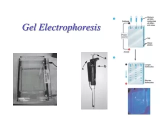
Background Agarose gel electrophoresis is the easiest and commonest way of separating and analyzing DNA. The purpose of the gel might be to look at the DNA, to quantify it or to isolate a particular band. The DNA is visualised in the gel by addition of ethidium bromide. This binds strongly to DNA by intercalating between the bases and is fluorescent meaning that it absorbs invisible UV light and transmits the energy as visible orange light.
What percentage gel ? Most agarose gels are made between 0.7% and 2%. A 0.7% gel will show good separation (resolution) of large DNA fragments (5–10kb) and a 2% gel will show good resolution for small fragments (0.2–1kb). Some people go as high as 3% for separating very tiny fragments but a vertical polyacrylamide gel is more appropriate in this case. Low percentage gels are very weak and may break when you try to lift them. High percentage gels are often brittle and do not set evenly. I usually make 1% gels. Which gel tank ? Small 8x10cm gels (minigels) are very popular and give good photographs. Larger gels are used for applications such as Southern and Northern blotting. The volume of agarose required for a minigel is around 30–50mL, for a larger gel it may be 250mL. This method assumes you are making a mini-gel.
How much DNA should I load ? The big question. You may be preparing an analytical gel to just look at your DNA. Alternatively, you may be preparing a preparative gel to separate a DNA fragment before cutting it out of the gel for further treatment. Either way you want to be able to see the DNA bands under UV light in an ethidium-bromide-stained gel. Typically, a band is easily visible if it contains about 20ng of DNA. Now consider an example. Suppose you are digesting a plasmid that comprises 3kb of vector and 2kb of insert. You are using EcoRI (a common restriction enzyme) and you expect to see three bands: the linearised vector (3kb), the 5' end of the insert (0.5kb) and the 3' end of the insert (1.5kb). In order to see the smallest band (0.5kb) you want it to contain at least 20ng of DNA. The smallest band is 1/10th the size of the uncut plasmid. Therefore you need to cut 10x20ng, that is 200ng of DNA (0.2µg). Then your three bands will contain 120ng, 20ng and 60ng of DNA respectively. All three bands will be clearly visible on the gel and the biggest band will be six times brighter than the smallest band.
How much DNA should I load ? Now imagine cutting the same plasmid with BamHI (another popular restriction enzyme) and that BamHI only cuts the plasmid once, to linearise it. If you digest 200ng of DNA in this case then the band will contain 200ng of DNA and will be very bright and will probably be overloaded. Too much DNA loaded onto a gel is a bad thing. The band appears to run fast (implying that it is smaller than it really is) and in extreme cases can mess up the electrical field for the other bands, making them appear the wrong size also. Too little DNA is only a problem in that you will not be able to see the smallest bands because they are too faint.Having said all that, I usually digest and load 2–4µL of the 50µL obtained from a kit miniprep. But you see how it depends on the number and size of the bands expected. For PCR reactions, it depends on the PCR but in routine applications 10–20µL should be plenty to see the product on the gel.
Which comb? This depends on the volume of DNA you are loading and the number of samples. Combs with many tiny teeth may hold 10µL. This is no good if you want to load 20µL of restriction digest plus 5µL of loading buffer. When deciding whether a comb has enough teeth, remember that you need to load at least one marker lane, preferably two. Making the gel (for a 1% gel, 50mL volume) Weigh out 0.5g of agarose into a 250mL conical flask. Add 50mL of 0.5xTBE , swirl to mix. It is good to use a large container, as long as it fits in the microwave, because the agarose boils over easily. Microwave for about 1 minute to dissolve the agarose. The agarose solution can boil over very easily so keep checking it. It is good to stop it after 45 seconds and give it a swirl. It can become superheated and NOT boil until you take it out whereupon it boils out all over you hands. So wear gloves and hold it at arms length. You can use a bunsen burner instead of a microwave - just remember to keep watching it. Leave it to cool on the bench for 5 minutes down to about 60°C (just too hot to keep holding in bare hands). If you had to boil it for a long time to dissolve the agarose then you may have lost some water to water-vapour. You can weigh the flask before and after heating and add in a little distilled water to make up this lost volume. While the agarose is cooling, prepare the gel tank ready, on a level surface.
Add 1µL of ethidium bromide (10mg/mL) and swirl to mix The reason for allowing the agarose to cool a little before this step is to minimise production of ethidium bromide vapour. Ethidium Bromide is mutagenic and should be handled with extreme caution. Dispose of the contaminated tip into a dedicated ethidium bromide waste container. 10mg/mLethidium bromide solution is made up using tablets (to avoid weighing out powder) and is stored at 4°C in the dark with TOXIC labels on it. Pour the gel slowly into the tank. Push any bubbles away to the side using a disposable tip. Insert the comb and double check that it is correctly positioned. The benefit of pouring slowly is that most bubbles stay up in the flask. Rinse out the flask immediately. Leave to set for at least 30 minutes, preferably 1 hour, with the lid on if possible. The gel may look set much sooner but running DNA into a gel too soon can give terrible-looking results with smeary diffuse bands. Pour 0.5x TBE buffer into the gel tank to submerge the gel to 2–5mm depth. This is the running buffer. You must use the same buffer at this stage as you used to make the gel. ie. If you used 0.6xTBE in the gel then use 0.6xTBE for the running buffer. Remember to remove the metal gel-formers if your gel tank uses them.
Preparing the samples Transfer an appropriate amount of each sample to a fresh microfuge tube. It may be 10µL of a 50µL PCR reaction or 5µL of a 20µL restriction enzyme digestion. If you are loading the entire 20µL of a 20µL PCR reaction or enzyme digestion (as I often do) then there is no need to use fresh tubes, just add in the loading buffer into the PCR tubes. Write in your lab-book the physical order of the tubes so you can identify the lanes on the gel photograph. Add an appropriate amount of loading buffer into each tube and leave the tip in the tube. Add 0.2 volumes of loading buffer, eg. 2µL into a 10µL sample. The tip will be used again to load the gel. Load the first well with marker.I store my markers ready-mixed with loading buffer at 4°C. I know to load 2µL and how much DNA is in each band. See below for more on this.Avoid using the end wells if possible. For example, If you have 12 samples and 2 markers then you will use 14 lanes in total. If your comb formed 18 wells then you will not be using 4 wells. It is best to not use the outer wells because they are the most likely to run aberrantly.
Continue loading the samples and finish of with a final lane of marker I load gels from right to left with the wells facing me. This is because gels are published, by convention, as if the wells were at the top and the DNA had run down the page. If this seems confusing then you can load left to right with the wells facing away from you. Close the gel tank, switch on the power-source and run the gel at 5V/cm. For example, if the electrodes are 10cm apart then run the gel at 50V. It is fine to run the gel slower than this but do not run any faster. Above 5V/cm the agarose may heat up and begin to melt with disastrous effects on your gel's resolution. Some people run the gel slowly at first (eg. 2V/cm for 10 minutes) to allow the DNA to move into the gel slowly and evenly, and then speed up the gel later. This may give better resolution. It is OK to run gels overnight at very low voltages, eg. 0.25–0.5V/cm, if you want to go home at 11 O'clock already.
Check that a current is flowing You can check this on the power-source, the milliamps should be in the same ball-park as the voltage, but the the best way is to look at the electrodes and check that they are evolving gas (ie. bubbles). If not then check the connections, that the power-source is plugged in etc.etc. This has been known to happen if people use water instead of running buffer. Monitor the progress of the gel by reference to the marker dye. Stop the gel when the bromophenol blue has run 3/4 the length of the gel. Switch off and unplug the gel tank and carry the gel (in its holder if possible) to the dark-room to look at on the UV light-box. Some gel holders are not UV transparent so you have to carefully place the gel onto the glass surface of the light-box. UV is carcinogenic and must not be allowed to shine on naked skin or eyes. So wear face protection, gloves and long sleeves.
Loading buffers The loading buffer gives colour and density to the sample to make it easy to load into the wells. Also, the dyes are negatively charged in neutral buffers and thus move in the same direction as the DNA during electrophoresis. This allows you to monitor the progress of the gel. The most common dyes are bromophenol blue (Sigma B8026) and xylene cyanol (Sigma X4126). Density is provided by glycerol or sucrose. Typical recipe- 25mg bromophenol blue or xylene cyanol - 4g sucrose - H2O to 10mL The exact amount of dye is not importantStore at 4°C to avoid mould growing in the sucrose. 10mL of loading buffer will last for years. Bromophenol blue migrates at a rate equivalent to 200–400bp DNA. If you want to see fragments anywhere near this size (ie. anything smaller than 600bp) then use the other dye because the bromophenol blue will obscure the visibility of the small fragments.Xylene cyanol migrates at approximately 4kb equivalence. So do not use this if you want to visualise fragments of 4kb.
Size markers There are lots of different kinds of DNA size markers. In the old days the cheapest defined DNA was from bacteriophage so alot of markers are phage DNA cut with restriction enzymes. Many of these are still very popular eg, lambda HindIII, lambda PstI, PhiX174 HaeIII. These give bands with known sizes but the sizes are arbitrary. Choose a marker with good resolution for the fragment size you expect to see in you sample lanes. For example, for tiny PCR products you might choose PhiX174 HaeIII but for 6kb fragments you would choose lambda HindIII. More recently, companies have started producing ladder markers with bands at defined intervals, eg. 0.5, 1, 1.5, 2, 2.5kb and so on up to 10kb. If you know the total amount of DNA loaded into a marker lane, and you know the sizes of all the bands, you can calculate the amount of DNA in each band visible on the gel. This can be very useful for quantifying the amount of DNA in your sample bands by comparison with the marker bands. It is good to load two markers lanes, flanking the samples. Lots of companies sell DNA size markers. It pays to shop around for the cheapest. Often the local kitchen-sink biotech company sells excellent markers.