Chapter 4 - Tissues
Chapter 4 - Tissues. Connective Tissue. Functions Binding and support Protection Insulation Transportation. Connective Tissue - Characteristics. Common Origin All arise from the mesenchyme (embryonic tissue) Degrees of vascularity
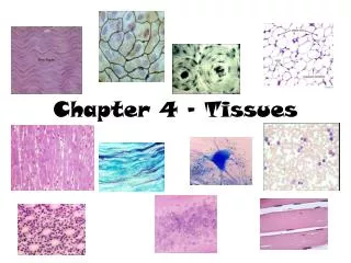

Chapter 4 - Tissues
E N D
Presentation Transcript
Connective Tissue • Functions • Binding and support • Protection • Insulation • Transportation
Connective Tissue - Characteristics • Common Origin • All arise from the mesenchyme (embryonic tissue) • Degrees of vascularity • Supply of blood vessels – vascularized to poorly vascularized. • Extracellular matrix • All other tissues are composed of cells. CT is composed mainly of the nonliving extracellular matrix which separates the living cells of the tissue. This allows it to bear weight, withstand tension, physical trauma, etc.
Connective Tissue • Structural elements • Ground Substance (matrix) • Fibers (matrix) • Cells
Connective Tissue • Ground Substance • Fills the space between the cells and contains the fibers. • Composed: • Interstitial fluid • Cell adhesion proteins • Glue to allow cells to attach themselves to matrix elements • Proteoglycans • Glycosaminoglycans (GAGs) attach and trap water to form anything from a fluid to a viscous gel – more GAGs, more viscous.
Connective Tissue - Fibers • Provide support • Types of fibers • Collagen fibers • “white fibers” has a white appearance • Very strong stronger than steel fibers! • Found in tendons and ligaments • Elastic fibers • “yellow fibers” has a yellow appearance • Able to stretch (protein elastin) – allows tissue to return to normal length and shape • Reticular fibers • Short, fine, collagenous fibers and are continuous with collagen fibers. • Reticul network • Often seen where connective tissue connects with other tissues forms a fuzzy “net” that allows more “give”
Connective Tissue - Cells • Each major class of c.t. has a fundamental cell. • Undifferentiated cells, indicated by –blast (“bud” “forming”) • Fibroblast – Connective tissue proper • Chondroblast – Cartilage • Osteoblast – Bone • Hematopoietic stem cell - Blood • Actively mitotic cells!!
Connective Tissue - Cells • Once the –blast cells synthesize the matrix, they assume their less active, mature mode, indicated by –cyte (fibrocyte, osteocyte….) • These cells maintain the health of the matrix! • If matrix is injured can revert back to –blast.
Connective Tissue - Cells • Other cells found within C.T. • White blood cells – defensive (neutrophils, eosinophils, lymphocytes) • Plasma cells – produces antibodies • Mast cells – oval, typically cluster along blood vessel walls. Detect foreign substances and initiate inflammatory response • Macrophages – large, irregularly shaped cells that phagocytize foreign materials. Found throughout loose C.T., bone marrow, and lymphatic tissue.
Connective Tissue • Most abundant and widely distributed tissue • Four main classes: • Connective tissue proper • Loose Connective Tissues • Areolar, adipose, and reticular • Dense Connective Tissues • Dense regular, dense irregular, elastic • Cartilage • Hyaline, elastic, fibrous • Bone Tissue • Blood
Connective TissueAreolar • Description • Gel-like matrix with all three fiber types; cells: fibroblasts, macrophages, mast cells and some wbc’s • Function • Wraps and cushions organs; its macrophages phagocytize bacteria; plays important role in inflammation; holds and conveys tissue fluid. • Location • Widely distributed under epithelia of body.
Connective TissueAdipose • Description • Fat!! • Accounts for about 18% of body weight • Function • Provides reserve food fuel; insulates against heat loss; supports and protects organs • Location • Under skin; around kidneys and eyeballs; within abdomen; in breasts
Connective TissueReticular • Description • Network of reticular fibers in a typical loose ground substance; reticular cells lie on the network. • Function • Fibers from a soft internal skeleton (stroma) that supports other cell types including wbc’s, mast cells, and macrophages. • Location • Lymphoid organs (lymph nodes, bone marrow, and spleen).
Connective TissueDense Regular • Description • Closely packed collagen fibers all running in the same direction • Poorly vascularized • Function • Attaches muscles to bones or to muscles; attaches bones to bones; withstands great tensile stress when pulling force is applied in one direction • Location • Tendons and ligaments
Connective TissueDense Irregular • Description • Thick collagen fibers that run in all directions • Function • Able to withstand tension exerted in many directions; provides structural strength • Location • Dermis of the skin; submucosa of digestive tract; fibrous capsules of organs and of joints
Connective TissueHyaline Cartilage • Description • Gristle • Most common • Amorphous but firm matrix; collagen fibers from and imperceptible network; chondroblasts produce the matrix and when mature (chondrocytes) lie in lacunae
Connective TissueHyaline Cartilage • Function • Supports and reinforces; has resilient cushioning properties; resists compressive stress • Location • Forms most of the embryonic skeleton; covers the ends of long bones in joint cavities; forms costal cartilages of the ribs; cartilages of the nose, trachea and larynx
Connective TissueElastic Cartilage • Description • Similar to hyaline cartilage, but more elastic fibers in the matrix • Function • Maintains the shape of a structure while allowing great flexibility • Location • Ear, tip of nose, epiglottis
Connective TissueFibrocartilage • Description • Found in areas of high stress • Matrix similar to but less firm that hyaline; thick collagen fibers predominate • Avascular • Function • Tensile strength with the ability to absorb compressive shock, prevents bone-to-bone contact, shock absorber • Location • Spinal discs, pads within knee joint
Connective TissueBone • Description • Hard, calcified matrix contain many collagen fibers; osteocytes lie in lacunae • Highly vascularized • Function • Supports and protects; levers for the muscles; stores calcium and other minerals and fat; marrow on inside make blood cells • Location • Bone
Connective TissueBlood • Description • Red and white blood cells in a fluid matrix (plasma) • Function • Transport respiratory gases, wastes, nutrients, immune response, and blood clotting • Location • Contained within blood vessels