Download
1 / 34
340 likes | 536 Vues
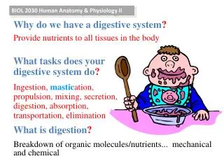
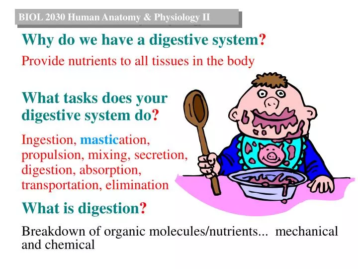
BIOL 2030 Human Anatomy & Physiology II. Why do we have a digestive system ? Provide nutrients to all tissues in the body. What tasks does your digestive system do ? Ingestion, mastic ation, propulsion, mixing, secretion, digestion, absorption, transportation, elimination.

E N D
BIOL 2030 Human Anatomy & Physiology II Why do we have a digestive system? Provide nutrients to all tissues in the body What tasks does your digestive system do? Ingestion, mastication, propulsion, mixing, secretion, digestion, absorption, transportation, elimination What is digestion? Breakdown of organic molecules/nutrients... mechanical and chemical
See Table 24.1 What happens between my mouth and anus? Organ Function Time ingestion/mastication 10-20 sec.secretion/mixing/digestion Oral cavity Pharynx Esophagus Stomach Small intestines Pancreas Liver Large intestines Rectum Anus transportation 1-2 sec. transportation/propulsion 5-8 sec. digestion/secretion 3-4 hrs.mixing digestion/secretion/mixing 3-5 hrs.absorption/propulsion absorption/propulsion 18-36 hrs.elimination
What structures exist between mouth and anus? Graphic overview of alimentary canal
What is the oral cavity and what does it do? Graphic overview of …
What is the oral cavity and what does it do? Vestibule: space between cheeks, lips and gums Oral cavity proper:space within floor of mouth and roof (palate) permanent deciduous Ingestion (alternate routes) Mastication = chewingWhich type of digestion? Why?Accomplished via: cheeks, lips, tongue, teeth, jaws Types of teeth:IncisorsCaninesPremolarsMolars
How do you masticate? Teeth embedded in:Alveoli in the Maxilla(upper jaw) & Mandible (lower jaw) Closing jaw =massetertemporalismedial pterygoids Opening jaw =lateral pterygoids
How do you salivate? 3 paired glands:ParotidSubmandibularSublingual Scattered small tubular glands Secrete mucus, serous fluid and enzymes Endocrine or Exocrine?
What can your palate do for you ? Hard and Soft palates Cleft lip/palate:congenital defect where embryonic tissues fail to fuse “Because I was different” by Don Bartlette http://smiletrain.org/
How does food get from oral cavity to esophagus? Food formed into a bolus Pushed against hard palate by tongue Initiates swallowing reflex Pharyngeal muscles aid in pushing into esophagus Swallowing (deglutition) divided into 3 phases:1) voluntary2) pharyngeal3) esophageal Which of the pharynxes usually conduct food?
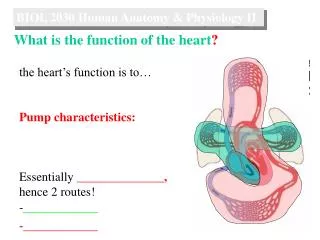
What is the role of the esophagus? Conducting Tube ~ 12” in length 2 sphincters:upper esophageal lower esophageal or cardiac Generalized layers of alimentary canal 4 1 2 3
What is the role of the enteric plexus? Nerve plexus allows local reflexes to be integrated independent of the CNS. Sensory neurons Motor neurons Interneurons 4 1 2 3
What’s unique about the stomique? Varies in size Rugae (wrinkles) allow distention Entrance = gastroesophageal opening (cardiac) Exit = pyloric opening Muscularis different because 3 layers of muscle DisordersHiatal hernia & Hypertrophic pyloric stenosis Endoscopy
How does gastric digestion occur? 5 types of cells: 1) Surface mucous cells = mucus (1-1.5 mm, alkaline) 2) Mucous neck cells = mucus 3) Parietal cells= HCl, Intrinsic factor (B12)
How does gastric digestion occur? 5 types of cells: 4) Chief cells = pepsinogen, becomes pepsin in low pH, breaks covalent protein bonds, making peptides 5) Endocrine cells = hormones (gastrin), which stimulates secretory activity of parietal cells. Gastric secretions and bolus form Chyme
How is gastric secretion regulated? Gastric secretion is divided into 3 phases: 1) Cephalic: Stimuli from head region initiate gastric secretion 2) Gastric: Stimuli from w/n stomach cause > secretion 3) Intestinal: Dependent on pH/chemistry of chyme in duodenum. > pH 3 then stimulates, < pH 2 then inhibits.
What is important about Liver anatomy? Largest internal organ, 2 major, 2 minor lobes Connective tissue capsule invades at hepatic portal, form membrane bound lobules (6-sided), portal triads in corners
What’s the digestive role of the Liver? Bile production: formed in lobules, drains out hepatic ducts, secreted and/or stored in gall bladder.Storage: can store nutrients (glycogen, vitamins, fats)Nutrient interconversion: metabolically “swapped” DetoxificationPhagocytosisSynthesis What are gall stones?
What’s the digestive role of the Pancreas? Exocrine and endocrine functions Pancreatic juice = Enzymatic component: enzymes essential for all food class digestion. Examples:trypsin = protein amylase = carboslipase = fats ribo and deoxyribonuclease = RNA/DNA
What’s the digestive role of the Pancreas? Pancreatic juice = Aqueous component H2O and HCO3-ions dilute & neutralize acidic chyme. What is a source of bicarbonate ions?
What are the regions of the small intestines? Long segment of alimentary canal… 6 meters 3 regions: 1) Duodenum 2) Jejunum 3) Ileum
What’s the internal anatomy of the small intestines? Contains features such as: plicae circularis (circular folds)villimicrovilli Why? Functions include: Digestion/Mixing/Absorption Digestive enzymes in association w/ microvilli: disaccharidases, peptidases, nucleases
What are the components of the colon? Divided into 4 regions:1) Ascending colonbegins at ileocecal valveends at hepatic flexure 2) Transverse colonbegins at hepatic flexureends at spleenic flexure 3) Descending colonbegins at spleenic flexureends at opening of pelvis Rectum 4) Sigmoid colonbegins at opening of pelvisends at rectum Sphincters Anus
What are the functions of the colon? SecretionAbsorption(form feces) Normal flora can produce: Vitamins (K)Flatus Capable of mass movements
How does it (defecation) happen? • Mass movements • Colic distension results in defecation reflex w/c is a contraction of rectal tunica muscularis and relaxation of internal anal sphincter. • If ignored reflex is extinguished. • If acted upon, voluntary actions result in defecation. Diarrhea & Constipation
What is the big picture of digestion? To understand Digestion and then absorption… You need to know the component “pieces” molecules of each major nutrient group. Water = H2O Vitamins/Minerals = B12, Ascorbic acid (C) etc. Salts = Positive ion (Ca2+) and negative ion (Cl-) CaCl2 Carbohydrates = Polysaccharides Disaccharides Monosac. Lipids = triglycerides 3 fatty acids & gylcerol Proteins = Polypeptides Dipeptides amino acids Bile Small intestine enzymes
What is the big picture of carbohydrate digestion? • Goal: Convert complex carbs (polysaccharides) and disacchrides into monosaccharides • Absorbed via 2ndary active transport • Facilitated diffusion to blood • Monosac. to liver to be converted to glucose and or glycogen Bile Small intestine enzymes
What is the big picture of lipid digestion? • Goal: Convert to fatty acids, glycerol • Micelles formed • Attach to plasma membrane, lipid components diffuse into epithelium • Changed back into triglycerides and coated with proteins. Chylomicrons excreted • Chylomicrons move to lacteal, then blood stream Bile Small intestine enzymes
What is the big picture of lipid digestion? What is the most dense? Water Lipids Proteins Bile Small intestine enzymes
What is the big picture of lipid digestion? Bile Small intestine enzymes
What is the big picture of protein digestion? • Goal: Form amino acids • Amino acids absorbed via 2ndary active transport and/or diffusion • A.a. moved out via active transport • Enter blood stream and move to liver Bile Small intestine enzymes
What is the big picture of protein digestion? Pepsin= polypeptides
What is the big picture of protein digestion? oligopeptides Trypsin= Chymotrypsin= Carboxypeptidase= oligopeptides amino acids
What is the big picture of protein digestion? amino acids Carboxypeptidase= Aminopeptidase= Dipeptidase= amino acids amino acids
Polysacc. Disacc. Poly, di peptides Disacc. Glycerol, fatty acids tri, di peptides, a. a. Monosacc. What is the big picture of digestion? Carbos Proteins Lipids Saliva amylase Gastric juice pepsin Bile bile salts Pancreatic juice trypsin lipase, esterase amylase Small intestine enzymes SucraseLactase peptidase lipase