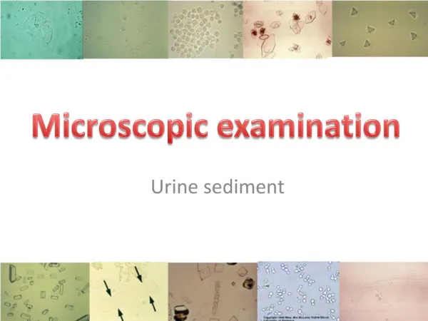
Urine Sediment Identification
Urine Sediment Identification. Penny S. Stevens, MBS, MT(ASCP), CLS(NCA) Sr. International QA/QC Medical Technologist SMILE, Johns Hopkins University. Course Objectives. Review the kidney structure Review rapid chemical tests Identify normal and abnormal urine sediment

Urine Sediment Identification
E N D
Presentation Transcript
Urine Sediment Identification Penny S. Stevens, MBS, MT(ASCP), CLS(NCA) Sr. International QA/QC Medical Technologist SMILE, Johns Hopkins University
Course Objectives • Review the kidney structure • Review rapid chemical tests • Identify normal and abnormal urine sediment • Review steps for accurate sediment ID • Case studies
Kidney Structure - Overview • Approximately 1 million nephrons in each kidney • Waste products and water pass along the tubule • Water, salts, sugar, & other large molecules reabsorbed within limits. • Excess products & Urine are excreted.
Bilirubin Blood Glucose Ketones pH Protein Urobilinogen Nitrite Leukocytes Specific Gravity Rapid Chemical Test
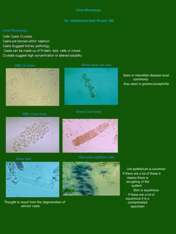
Normal & Abnormal Microscopy Review • Cells • Casts • Crystals Uric Acid Crystals RBC’s & WBC’s Epithelial Cast
Microscopy - Cells • Red Blood Cell • What rapid chemistry result do you expect to be positive? • Blood • Leukocyte Esterase • Possibly Nitrite
Microscopy - Cells • White Blood Cell • What rapid chemistry result do you expect to be positive? • Leukocyte Esterase
Microscopy - Cells • Squamous Epithelial • What rapid chemistry result do you expect to be positive? • Possibly Nitrite
Microscopy - Cells • Renal Epithelial • What rapid chemistry result do you expect to be positive? • Possibly Protein
Kidney Structure – Cast Formation • Protein – Tamm-Horsfall • Secreted by tubule cells • Distal convoluted tubule (DCT). • T-H Protein, albumin & immunoglobulin • Combine to form cast • Include tubule contents • Illustration: • Protein secretion (green dots) • Hyaline cast (formed in the collecting duct.
Cast Formation • DST protein secretion • Tubule contents wrapped into protein matrix • None = Hyaline • Cells = Cellular Cast • Progression • Cellular • Coarse Granular • Fine Granular • Waxy – (Theory)
Microscopy – Cast ID • Hyaline Cast • Key Features • Transparent • Cylindrical • Rounded Ends • Colorless • Homogenous • Nearly Parallel sides • No Dark Edges
Microscopy – Cast ID • Hyaline Cast • What rapid chemistry result do you expect to be positive? • Protein: • Negative – 1+
Microscopy – Cast ID • Cellular Cast – (Renal) • Key Features • Cellular: Few – Numerous • Cylindrical • Rounded Ends • Color – depends on the contents • Nearly Parallel sides • No Dark Edges
Microscopy – Cast ID • Fatty Cast – Polarized Light • Maltese cross • Similar to starch • What rapid chemistry result do you expect to be positive? • Protein • Other rapid chemistries- Depends on the contents Starch - Polarized Fat - Polarized
Microscopy – Cast ID • Granular Cast • Coarse - black • Fine – grey/pale yellow • Key Features • Cellularity Indeterminate • Cylindrical • Rounded Ends • Nearly Parallel sides • No Dark Edges Coarse Granular Cast Fine Granular Cast
Microscopy – Cast ID • Granular Cast (Hemoglobin) • What rapid chemistry result do you expect to be positive? • Protein • Other - Depends on the origin: • Blood/Hemoglobin
Microscopy – Cast ID • Waxy Cast • Key Features • Opaque (Refractive) • Homogeneous (non-cellular) • At Least One Flat/Blunt End • Short • Possibly Cracked, Serrated or Convoluted • Nearly Parallel Sides
Microscopy – Cast ID • Waxy Cast • What rapid chemistry result do you expect to be positive? • Protein • >3+
Microscopy – Artifact • Fibers • Key Features • Refractile • Flat • Parallel Sides • Dark Edges
Cast Comparison • Opaque • Cylindrical • Blunt Ends Hyaline Cast Waxy Cast Fiber • Transparent • Cylindrical • Rounded Ends • Refractile • Flat • Dark Edges
Urine Sediment Training • Questions??