Integument System Chapter 6
Integument System Chapter 6. Our largest external organ!!. Skin? . The Integumentary system is a remarkable structure that is fundamentally integrated with the other organ systems. It is the largest human organ system .

Integument System Chapter 6
E N D
Presentation Transcript
Integument SystemChapter 6 Our largest external organ!!
Skin? • The Integumentary system is a remarkable structure that is fundamentally integrated with the other organ systems. • It is the largest human organ system. • Developmental defects of the Integumentary system compromise the ability of other organ systems to maintain the body’s homeostasis. • The skin’s ability to lose or retain body heat plays a major role in regulating body temperature. • Skin damage and disease compromise the skin’s ability to do its work.
Skin Functions • Skin provides four functions for the body: • protection • heat regulation • sensation • waste excretion • Helps to maintain body homeostasis
Skin Contents • Blood vessels • Connective tissue • Glands • Hair • Nails • Skin
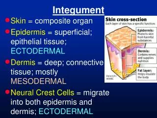
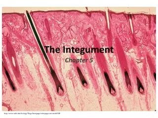
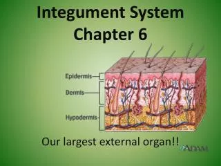
Skin Structure • Skin is composed of three distinct layers: • epidermis • dermis • subcutaneous layer or hypodermis
Skin • Epidermis • composed of stratified squamous epithelium • 5 or 6 layers • The upper layers of skin fill with keratin and die. • Dermal-Epidermal Junction • Polysaccharide gel “glues” the two layer together • Large areas of detachment can lead to infection
Layers of the Epidermis • Stratum Corneum • Horny layer • Outermost layer has water resistant protein (keratin) • Adjacent cells held together by desmosomes • Stratum Lucidum • Clear layer • Filled with gel-like substance called eleidin • Eleidin blocks water loss and water penetration
Layers of Epidermis • Stratum Granulosum • Granular layer • Keratinization begins here • Cells in this layer begin to degenerate • Stratum Spinosum • Spiny layer • Irregularly shaped cells with prominent bridges or desmosomes • Cells rich in RNA, well equiped to initiate protein synthesis for Keratin
Layers of Epidermis • Stratum Basale • Base layer • Single layer of columnar cells • Only layer to undergo mitosis • Calluses form due to an over production of skin cells and lack of shedding
Epidermal Cell Types • Keratinocytes • Filled with tough fibrous protein Keratin • Make up 90% of the cells • Old cells are shed every 35-45 days • Melanocytes • Gives color to the skin (can be completely absent) • Decreases amount of UV light
Epidermal Cell Types • Langerhans cells • Play limited role in immune responses • Serve as defense mechanism • Originate in bone marrow and migrate to the epidermis early • Work with helper T cells (WBC) to trigger immune response
Skin Structure (Dermis) • The dermis is primarily dense irregular connective tissue. • Three types of fibers are found in the dermis: • collagen • elastin • reticulin • The dermis contains nerves and blood vessels. • The subcutaneous layer contains adipose tissue.
Dermis • Composed of 2 layers Papillary & Reticular • Thicker than the epidermis • Contains most or all of the mechanical strength • Reservoir storage for H2O and Electrolytes
Dermis • Papillary Layer • Location of most of the blood vessels • Dermal papillae • Bumps that project into epidermis (prints) • Allows us to grip surfaces and walk upright • Reticular Layer • Tough and elastic • Attachment for smooth and skeletal muscle • Arrectorpili muscle • Goose bumps
Skin Color • Melanocytes in the stratum basale is about the same in all races… • Why the color difference?? • The amount of melanin produced varies • Melanocyte • Produces pigment • Converts the amino acid tyrosine into the dark brown melanin pigment • Pigment is transferred to other epidermal cells
This Pigment Process • Dependent on many factors • Genetics/heredity (4-6 pairs of genes) • Exposure to sunlight • Age • The absence of pigment Albinism • Color is also absent from iris of the eye and the hair • Regulated by tyrosinase • Color changes affected by blood flow to the skin 18
ABCD of Moles • A – asymmetry • Is it lopsided • B – border • Edges indistinct • C – color • Uneven or mixture of shades • D – diameter • Larger than ¼ of an inch 19
Role of our Skin • Protection • Sensation • Permits movement and growth • Excretion • Vitamin D production • Immunity • Homeostasis of body temperature 21
Heat Loss and Burns • 80% of our heat loss occurs through the skin • Evaporation • Water on surface leaves • Radiation • Microwaves • Conduction • Direct contact with surface • Convection • Circulating air • Body surface areas • The larger the area burned, the less chance of survival • Rule of Nines • Body is divided into 11 areas of 9% with the genitals being 1%
Burns • Burns are categorized according to severity of skin damage: • First-degree burn • sunburn • Second-degree burn • Deep epidermal layers; scarring • Third-degree burn • A full thickness burn • Destruction of epidermis and dermis • May involve muscle and bone • Insensitive to pain immediately after the injury due to nerve destruction
Skin Appendages • The skin contains several types of glands. • Nerves provide the skin with sensation. • Nails are sheets of dried, flattened, keratinized cells. • Hair is composed of cylinders of keratinized cells.
Hair • Found over all areas of the body, except palms and soles of feet • Lanugo • Fine baby hair you are born with • Replaced with Vellus hair (stronger, finer, less pigment) • Terminal hair pubic and axillary areas
Hair • Hair growth begins when the cells of the epidermis spread to the dermis forming small tubes called follicles • Average growth = ½ inch per month • Not stimulated by cutting or shaving • Sebaceous glands located at base of the follicle to provide lubrication
Nails • Made of flattened keratin cells
Skin glands • Sweat glands • Apocrine • Found in limited areas (arm pits) • Begin to function at puberty • Eccrine • Most numerous, help maintain core body temp • Hands have about 3000
Skin glands • Oil glands • Secrete sebum (nature’s skin cream) • Keeps hair soft and prevents water loss • Lipid components have and antifungal property • Ceruminous glands • Modified apocrine sweat glands • Empty contents into external ear canal • May be mixed with sebaceous fluid • Excess secretions can cause blockages and hearing loss
Skin Infections • Impetigo • Bacterial, highly contagious, blisters with yellow crust • Tinea • Fungal infections (ring worm, jock itch, athlete’s foot) • Warts • Papillomavirus, can be contagious and cancerous • Boils • Local staphylococcus infection of the hair follicle
Skin Cancer • Basal cell carcinoma • the least malignant (cancerous; spreads throughout the body) type • the most common type • removal of the cancer area by surgery cures 99% of all cases • Squamous cell carcinoma • starts in the keratinocytes of the stratum spinosum • surgical removal and radiation therapy cures most cases • Malignant melanoma • cancer of the melanocytes • the most dangerous type • accounts for 5% of skin cancers • 1/3 of cases develop from pigmented moles
Aging • Some of skin aging is due to intrinsic factors. • Some of skin aging is due to extrinsic, or environmental, factors. • Lifestyle may accelerate skin aging. • Give some examples THE END