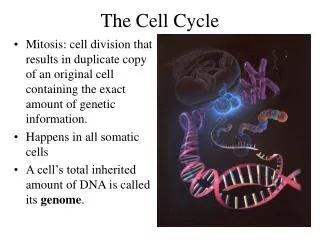
The Cell Cycle
The Cell Cycle. Pages 1067-1071, 1087-1088, 1105-1107. S phase. -Functions -Duplicate the DNA -Protein packing must be reproduced -2 issues facing DNA replication -Accuracy of replication -Copy DNA only once. Control of DNA Duplication. 2 steps in Replication

The Cell Cycle
E N D
Presentation Transcript
The Cell Cycle Pages 1067-1071, 1087-1088, 1105-1107
S phase -Functions -Duplicate the DNA -Protein packing must be reproduced -2 issues facing DNA replication -Accuracy of replication -Copy DNA only once
Control of DNA Duplication 2 steps in Replication 1. Late M/Early G1 -Formation of prereplicative complex (pre-RC) 2. Start of S phase -Preinitiation complex
Initiation of DNA Replication ORCs are always bound to the origins pre-RC assembly: -Inhibited by Cdk activity -Stimulated by APC/C
Initiation of DNA Replication -S-Cdk triggers the preinitiation complex -Diassembly of pre-RC -ORC and Cdc6 is P by Cdk -Cdc6 is degraded -Cdt1 is inhibited by geminin
Initiation of DNA Replication -DNA Replication is completed -In late M, APC/C the degradation of geminin -Cdk activity is decreased, OCRs are dephosphorylated -Cdc6 is synthesized -pre-RC assembly
Duplication of Chromatin Structure -Cdks stimulate synthesis of the 4 histone subunits -Nucleosome assembly factors typically associate with the replication fork -Histone modifying enzymes are thought to play a role
Cohesins and Sister Chromatids -Cohesin – large protein complex which holds the sister chromatids together Smc (Structural Maintenance of Chromosomes)
P Sister Chromatid Separation Securin inhibits separase Separase cleaves cohesin
DNA DamageCheckpoint Mdm2 – ubiquitin ligase p21 – CKI (Cdk Inhibitor Protein)
Check1/2 Chk1/2 phosphorylate Cdc25 thereby inactivating the phosphatase activity resulting in inactive Cdk
DNA Damage -Cell cycle is arrested until the damage is repaired -If it’s not repaired: 1. Unicellular organisms will resume their cycle taking a potential mutation over death 2. Multicellular organimsms will sacrifice a cell over the health of the organism
Apoptosis Pages 1115-1128
Apoptosis Apoptosis is one type of programmed Process of Apoptosis -Cells shrink and condense -Cytoskeleton collapses -Nuclear envelope disassembles -Nuclear chromatin fragments -Cell surface belbs and may break up (apoptotic bodies) -Cell surface is chemically altered -Macrophage engulfs the cell Process of Cell Necrosis -Cell insult or injury -Cells swell and burst -Cell contents are released -Inflammatory response
Examples of Apoptosis 1. Number of cells -Nervous system
Examples of Apoptosis 2. Development Paws of embryonic mouse
Examples of Apoptosis 3. Regulate cell numbers -Liver 4. Quality control -Eliminates abnormal, misplaced, nonfunctional, or dangerous cells -Developing T and B cells that do not produce useful antigen receptors or that are self-reactive 5. Supply of cells -Large numbers of neutrophils are produced and stored awaiting infection
Biochemical Characteristics DNA Cleavage -Endonuclease cleaves DNA into fragments between nucleosomes
Biochemical Characteristics TUNEL Assay - TdT-mediated dUTP nick end labeling Phosphatidylserine localization -movement from the inner to outer membrane -marks the cell for macrophages
Caspases -Enzymes responsible for apoptosis -A family of proteases that cleave proteins at aspartic acid residues -C for cysteine (in active site) and ASP for aspartic acid -Synthesized as inactive precursors, procaspases -Not all caspases are involved with apoptosis -ICE, interleukin-1-converting enzyme
Caspases -Initiator procaspase, start the proteolytic cascade -Executioner procaspases, cleave and activate other executioner procaspases and other targets -Targets include: nuclear lamins, endonuclease inhibitor, cytoskeleton components, cell-cell adhesion proteins -The caspase cascade is: -Very destructive -Self-amplyfing -Irreversible
Caspases -Initiator caspases have a caspase recruitment domain (CARD) that enables them to bind to adaptor proteins into activation complexes -In the complex, the initiator caspases are close enough to activate each other starting the cascade -2 Apoptotic pathways 1. Extrinsic Pathway 2. Intrinsic Pathway
Extrinsic Pathway DISC – death-inducing signaling complex Inhibitors such as decoy receptors and intracellular blocking proteins
Intrinsic Pathway Releases mitochondrial proteins into the cytoplasm Cytochrome C release can trigger apoptosis through interaction with the adapter protein Apaf1
Classes of Bcl2 Proteins Bcl2 proteins –regulate apoptosis through controlling the release of cytochrome c
Regulation of Intrinsic Pathway BH3-only proteins activate apoptosis through direct binding with anti-apoptotic proteins -p53 activates BH3-only proteins (Puma and Noxa) -Bid – extrinsic and intrinsic pathways
Inhibition of Apoptosis (BH3-only pro-apoptotic) (Anti-apoptotic)