Download
1 / 24
240 likes | 422 Vues
Chapter 13 The Heart and Heart Disease. Location, Size, and Position of the Heart. Triangular organ located in mediastinum with two thirds of the mass to the left of the body midline and one third to the right; the apex is on the diaphragm; shape and size of a closed fist.

E N D
Location, Size, and Position of the Heart • Triangular organ located in mediastinum with two thirds of the mass to the left of the body midline and one third to the right; the apex is on the diaphragm; shape and size of a closed fist
Location, Size, and Position of the Heart • Cardiopulmonary resuscitation (CPR)—heart lies between the sternum in front and the bodies of the thoracic vertebrae behind; rhythmic compression of the heart between the sternum and vertebrae can maintain blood flow during cardiac arrest; if combined with artificial respiration procedure, it can be life saving
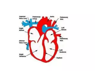
Anatomy of the Heart • Heart chambers • Two upper chambers are called atria (receiving chambers)—right and left atria • Two lower chambers called ventricles (discharging chambers)—right and left ventricles
Anatomy of the Heart • Wall of each heart chamber is composed of cardiac muscle tissue called myocardium • Endocardium—smooth lining of heart chambers—inflammation of endocardium called endocarditis
Anatomy of the Heart • The pericardium and pericarditis • Pericardium is a two-layered fibrous sac with a lubricated space between the two layers • Inner layer is called visceral pericardium or epicardium • Outer layer called parietal pericardium • Pericarditis—inflammation of the pericardium • Cardiac tamponade—compression of the heart caused by fluid building up between the visceral pericardium and parietal pericardium
Anatomy of the Heart • Heart action • Contraction of the heart is called systole; relaxation is called diastole • Heart valves and valve disorders
Anatomy of the Heart • Four valves keep blood flowing through the heart; prevent backflow (two atrioventricular or AV and two semilunar valves) • Tricuspid: at the opening of the right atrium into the ventricle • Bicuspid (mitral): at the opening of the left atrium into the ventricle
Anatomy of the Heart • Pulmonary semilunar: at the beginning of the pulmonary artery • Aortic semilunar: at the beginning of the aorta • Incompetent valves “leak,” allowing some blood back into the chamber from which it came
Anatomy of the Heart • Stenosed valves are narrower that normal, reducing blood flow • Rheumatic heart disease—cardiac damage resulting from a delayed inflammatory response to streptococcal infection • Mitral valve prolapse (MVP)—incompetence of mitral valve because its edges extend into the left atrium when the left ventricle contracts
Heart Sounds • Two distinct heart sounds in every heartbeat or cycle—“lubb-dupp” • First (lubb) sound is caused by the vibration and closure of AV valves during contraction of the ventricles • Second (dupp) sound is caused by the closure of the semilunar valves during relaxation of the ventricles • Heart murmurs—abnormal heart sounds often caused by abnormal valves
Blood Flow Through The Heart • Heart acts as two separate pumps—the right atrium and ventricle performing different functions from the left atrium and ventricle
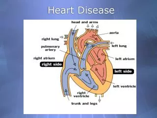
Blood Flow Through The Heart • Sequence of blood flow: venous blood enters the right atrium through the superior and inferior venae cavae—passes from the right atrium through the tricuspid valve to the right ventricle; from the right ventricle through the pulmonary semilunar valve to the pulmonary artery to the lungs—blood from the lungs to the left atrium, passing through the bicuspid (mitral) valve to left ventricle; blood in the left ventricle is pumped through the aortic semilunar valve into the aorta and is distributed to the body as a whole
Coronary Circulation and Coronary Heart Disease • Blood, which supplies oxygen and nutrients to the myocardium of the heart, flows through the right and left coronary arteries • Blockage of blood flow through the coronary arteries can cause myocardial infarction (heart attack)
Coronary Circulation and Coronary Heart Disease • Atherosclerosis (type of “hardening of arteries” in which lipids build up on the inside wall of blood vessels) can partially or totally block coronary blood flow • Angina pectoris—chest pain caused by inadequate oxygen to the heart
Cardiac Cycle • Heart beat is regular and rhythmic—each complete beat called a cardiac cycle—average is about 72 beats per minute • Each cycle, about 0.8 seconds long, subdivided into systole (contraction phase) and diastole (relaxation phase)
Cardiac Cycle • Stroke volume is the volume of blood ejected from one ventricle with each beat • Cardiac output is amount of blood that one ventricle can pump each minute—average is about 5 L per minute at rest
Conduction System of the Heart • Normal structure and function • SA (sinoatrial) node, the pacemaker—located in the wall of the right atrium near the opening of the superior vena cava • AV (atrioventricular) node—located in the right atrium along the lower part of the interatrial septum • AV bundle (bundle of His)—located in the septum of the ventricle • Purkinje fibers—located in the walls of the ventricles
Conduction System of the Heart • Electrocardiography • Specialized conduction system structures generate and transmit the electrical impulses that result in contraction of the heart • These tiny electrical impulses can be picked up on the surface of the body and transformed into visible tracings by a machine called an electrocardiograph • The visible tracing of these electrical signals is called an electrocardiogram or ECG
Conduction System of the Heart • The normal ECG has three deflections or waves called the P wave, the QRS complex, and the T wave • P wave—associated with depolarization of the atria • QRS complex—associated with depolarization of the ventricles • T wave—associated with repolarization of the ventricles
Conduction System of the Heart • Cardiac dysrhythmia—abnormality of heart rhythm • Heart block—conduction of impulses is blocked • Complete heart block—impaired AV node conduction, producing complete dissociation of P waves from QRS complexes • Can be treated by implanting an artificial pacemaker
Conduction System of the Heart • Bradycardia—slow heart rate (under 60 beats/min) • Tachycardia—rapid heart rate (over 100 beats/min) • Sinus dysrhythmia—variation in heart rate during breathing cycle • Premature contraction (extrasystole)—contraction that occurs sooner than expected in a normal rhythm • Fibrillation—condition in which cardiac muscle fibers are “out of step,” producing no effective pumping action
Heart Failure • Heart failure—inability to pump enough returned blood to sustain life; it can be caused by many different heart diseases • Right-sided heart failure—failure of the right side of the heart to pump blood, usually because the left side of the heart is not pumping effectively
Heart Failure • Left-sided heart failure (congestive heart failure)—inability of the left ventricle to pump effectively, resulting in congestion of the systemic and pulmonary circulations • Diseased hearts can be replaced by donated living hearts (transplants) or by artificial hearts (implants), although both procedures have yet to be perfected